
Short answer
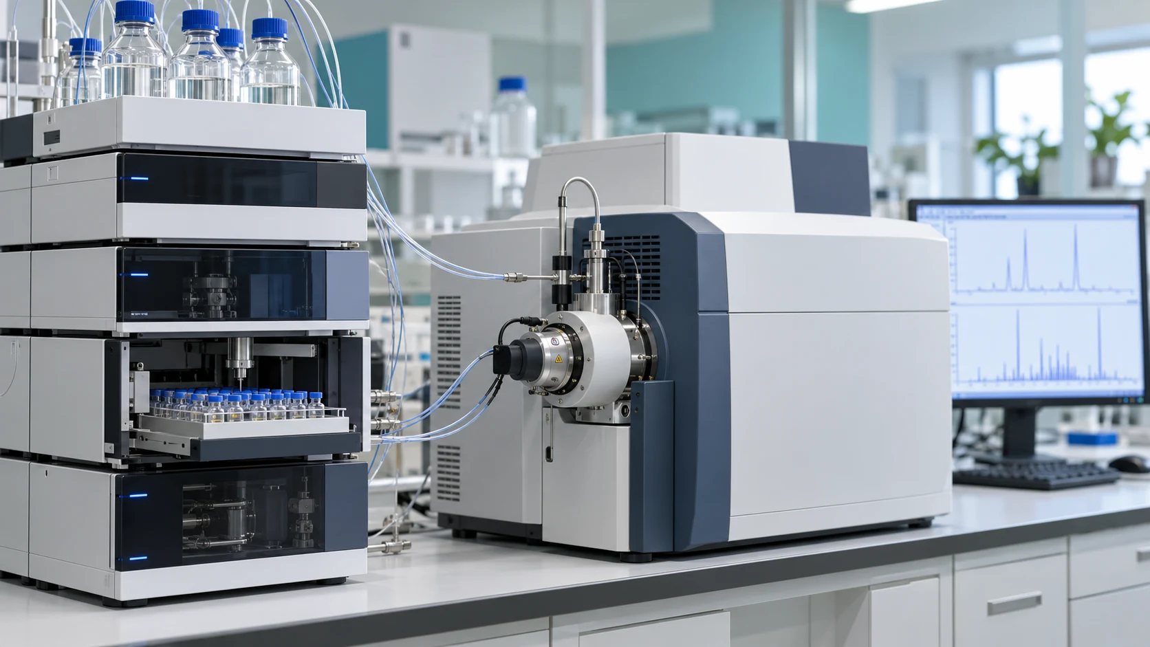
Peptide mapping techniques cut a protein into predictable peptides, separate them, and identify each one so the protein’s sequence and modifications can be checked. The field began in the 1950s with two-dimensional paper “fingerprints,” moved to HPLC and low-resolution mass spectrometry in the 1980s, and today relies on UHPLC coupled to high-resolution tandem mass spectrometry, supported by automated digestion and software that reports sequence coverage and post-translational modifications (PTMs).
For how a basic peptide map is run and read, see peptide mapping analysis for reliable research verification. This article covers how the techniques developed and how to choose among them.
What Are Peptide Mapping Techniques and Why Are They Important?
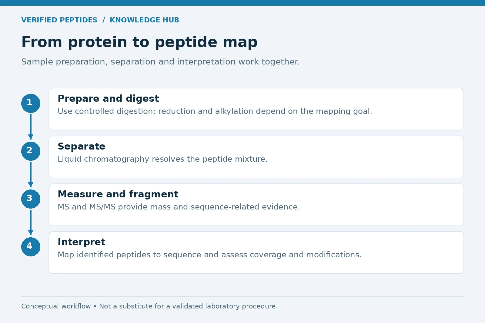
Peptide mapping techniques are analytical methods that break a protein into smaller peptides, separate those peptides, and identify them, producing a pattern that works like a fingerprint for that protein. Trypsin is the usual enzyme for the digestion step because of its high activity and cleavage specificity.1
It matters because one experiment can confirm identity and primary structure, show disulfide linkages, and reveal post-translational modifications (PTMs) such as site-specific glycosylation, along with sequence variants and truncations.2 Short synthetic peptides are a different case: they need no digestion, and suppliers typically document them with mass spectrometry and HPLC to establish composition and purity.3 How sequence data identify a peptide is covered in the role of amino acid sequencing in peptide identification.
A Historical Overview: How Peptide Mapping Techniques Have Evolved
The field moved from paper fingerprints in the 1950s, to HPLC with low-resolution mass spectrometry in the 1980s, to today’s UHPLC with high-resolution tandem mass spectrometry.
In 1956, Vernon Ingram used a two-step “fingerprinting” process, paper electrophoresis followed by paper chromatography, to compare normal hemoglobin with a variant hemoglobin (HbS). One spot differed between the two fingerprints, which pointed to a small difference in amino acid sequence.4, 5 Comparing digests instead of sequencing whole proteins still underpins the method today.
By the late 1980s and early 1990s, when the first monoclonal antibodies were mapped, HPLC columns were packed with 5 to 10 µm particles and run at 400 bar. Fast atom bombardment fed peptides into low-resolution mass spectrometers, and identities were confirmed by Edman degradation (stepwise chemical sequencing) after collecting peaks.6 Soft ionization changed what mass spectrometry could measure: electrospray ionization, published by John Fenn in 1988, and the laser desorption method Koichi Tanaka introduced around the same time made it possible to analyze large biological molecules, work recognized by the 2002 Nobel Prize in Chemistry.7
| Era | Separation | Detection and identification |
|---|---|---|
| 1950s | Paper electrophoresis, then paper chromatography5 | Visual spot pattern (“fingerprint”) |
| Late 1980s to early 1990s | HPLC, 5 to 10 µm particles, 400 bar6 | UV, low-resolution MS by fast atom bombardment, Edman degradation |
| 2000s | UPLC with 1.7 µm particles8 | Electrospray LC-MS with accurate mass |
| Today | UHPLC with sub-2 µm particles up to 1500 bar; micro-pillar array columns6 | High-resolution tandem MS, ion mobility, multi-attribute methods9, 10 |
The Role of Mass Spectrometry in Modern Peptide Mapping
Mass spectrometry advanced peptide mapping by replacing slow chemical sequencing with direct, accurate mass measurement and fragmentation of every peptide in a run. In LC-MS, liquid chromatography separates the digest and the mass spectrometer measures each peptide; in tandem mass spectrometry (MS/MS), selected peptide ions are broken apart in a sequence-informative way, so the fragments reveal the sequence and the site of any modification.11 Typical fragment patterns are explained in peptide fragmentation patterns in mass spectrometry.
Mass accuracy is a large part of the gain. In a 2006 Waters application note using 1.7 µm UPLC columns, the root-mean-square mass error across 35 selected peptides was 2.9 ppm (parts per million).8 Fragmentation options have widened too. Collision-based fragmentation often strips labile groups such as phosphate before the peptide backbone breaks; electron transfer dissociation (ETD), developed in the Hunt laboratory in the mid-2000s, has had its broadest impact on difficult-to-sequence PTMs such as phosphorylation and glycosylation.11
Peptide Mapping in Proteomics: Understanding Protein Structure and Function
In proteomics, the same digest, separate, and identify logic is scaled from one protein to thousands, so mapping becomes a way to study protein structure and function across whole samples. Mass spectrometry-based proteomics can identify and, increasingly, precisely quantify thousands of proteins from complex samples.12
For a single protein, peptide-level data show which regions carry modifications, which cysteines are linked by disulfide bonds, and whether the sequence matches what was expected.2
Large-scale proteomics and biomarker research
Peptide mapping supports large-scale proteomics and biomarker research by letting researchers compare peptide and protein patterns across many samples. Reviews of the field describe a pipeline that runs from sample preparation and separation through peptide fragmentation, PTM analysis, quantification, and bioinformatics, ending in biomarker discovery and validation.13 In pharmaceutical discovery research, the same tools give proof of identity and primary structural characterization for candidate proteins.2
Current State-of-the-Art Peptide Mapping Techniques
The most commonly used peptide mapping techniques in modern laboratories are enzymatic digestion followed by reversed-phase liquid chromatography with UV detection, LC-MS, or LC-MS/MS on high-resolution instruments, often called high-resolution mapping. Current columns use sub-2 µm porous or superficially porous (solid-core) particles at pressures up to 1500 bar.6
- Ion mobility, which adds a collision cross section (CCS) value to every peptide and simplifies spectra for low-abundance peptides that co-elute with abundant ones.9
- Multi-attribute methods (MAM), which use high-resolution mapping data to identify, quantify, and monitor several quality attributes at once.10
- Processing software that matches peptides to the sequence and reports sequence coverage, the share of the protein actually observed.14
Reversed-phase separation remains the workhorse for peptide maps; other modes are compared in an overview of HPLC modes for peptide separation.
How Peptide Mapping Techniques Are Used in Quality Control
In biopharmaceutical quality control, peptide maps confirm that each lot has the intended primary structure. ICH Q6B, the international guideline on specifications for biotechnological and biological products, describes peptide mapping with an appropriately validated procedure as a method frequently used to confirm desired product structure for lot release.15
Q6B adds that mapping under reducing and non-reducing conditions may help evaluate disulfide bridges, and that mapping may help detect some product-related variants.15 In the United States Pharmacopeia, general chapter <1055> on peptide mapping, harmonized with other pharmacopeias through the Pharmacopeial Discussion Group, was revised with its scope limited to confirmation of identity, and the revision became official on December 1, 2024.16
Mass spectrometry is often used alongside conventional LC-UV peptide maps.8 MAM goes further for biopharmaceuticals: it can reduce the number of separate tests for specific quality attributes, such as ion-exchange and capillary electrophoresis methods, and adds new peak detection, which flags unexpected peaks by comparison with a reference sample.10
Key Challenges in Peptide Mapping and How to Overcome Them
The main challenges are non-specific or incomplete digestion, artifacts created during sample preparation, and variation between runs and analysts. Each can be reduced with higher-specificity enzymes, shorter controlled digestions, automation, and replicate testing.
| Challenge | What happens | How labs address it |
|---|---|---|
| Non-specific cleavage | Some MS-grade trypsins showed chymotryptic-like activity and autolysis, creating extra peptides1 | Recombinant trypsin or a trypsin/Lys-C mix with high cleavage specificity1 |
| Incomplete digestion | Proteolytically resistant sites stay uncut, leaving gaps in the map1 | Higher enzyme amounts, possible when the enzyme is highly specific1 |
| Prep-induced modifications | Deamidation and oxidation created during sample preparation distort PTM results14 | Shorter, controlled digestions and method comparisons14 |
| Run-to-run variation | Lengthy manual preparation adds sample-to-sample variation14 | Automated reduction, alkylation, digestion, and desalting; replicate preparations14, 17 |
| Data complexity | Low-abundance peptides co-elute with abundant ones | Ion mobility and high-resolution MS for cleaner spectra9 |
Accuracy and reproducibility have to be demonstrated, not assumed. In a Thermo Fisher comparison, each digestion method was run as six preparations by two analysts over three days; an in-solution heat digestion produced about 10.8% oxidation at one methionine site in a monoclonal antibody, compared with about 3.3% to 3.8% for the other methods at the same site.14
Emerging Technologies Shaping the Future of Peptide Mapping
Machine learning, chip-based chromatography, ion mobility, and end-to-end automation are the technologies most likely to shape the next stage of the field. Several are already in use.
Machine learning in proteomics is now practical. Prosit, a deep learning model built on a synthetic peptide library extended to 550,000 tryptic peptides, predicts retention times and fragment ion intensities; adding it to database searches gave more identifications at more than tenfold lower false discovery rates (the estimated share of incorrect identifications).18
Micro-pillar array columns are made by lithographic etching, which creates an ordered separation bed on a silicon chip; efficiencies of up to 200,000 theoretical plates (a standard measure of column efficiency) within 30 minutes have been reported for peptide maps.6
How to Choose the Right Peptide Mapping Technique for Your Laboratory Needs
The right technique depends first on the question: routine identity, full characterization, PTM tracking, or monitoring several attributes at once. Match the method to the answer you need, then to your instruments, sample amount, and validation needs.
| Need | Common technique | Notes |
|---|---|---|
| Routine identity against a reference | LC-UV peptide map | Often paired with MS when peaks need to be named8 |
| Sequence confirmation and PTM sites | LC-MS/MS on a high-resolution instrument | ETD helps with labile PTMs11 |
| Disulfide bond assignment | Maps under reducing and non-reducing conditions | Described in ICH Q6B15 |
| Several attributes in one assay | MAM with new peak detection | Can reduce the number of separate tests10 |
| Short synthetic peptide | Intact mass by MS plus HPLC purity | Digestion is usually unnecessary3 |
For how HPLC data support characterization, see the role of HPLC analysis in peptide characterization.
Frequently Asked Questions About Peptide Mapping
How has mass spectrometry advanced peptide mapping techniques?
It replaced Edman sequencing of collected peaks with accurate mass measurement and MS/MS fragmentation of each peptide, so sequence and modification sites can be read within one LC-MS run.
What role does peptide mapping play in proteomics and pharmaceutical discovery research?
It supplies the peptide-level identifications that proteomics scales up to thousands of proteins, and in discovery research it helps confirm that candidate proteins carry the intended sequence and modifications.
How do peptide mapping techniques contribute to the quality control of biopharmaceuticals?
They confirm lot identity and primary structure, a use ICH Q6B describes for lot release, and they help track attributes such as oxidation, deamidation, and disulfide bonds.
What are the challenges in peptide mapping, and how can they be addressed?
The main ones are non-specific or incomplete digestion, artifacts from sample preparation, and run-to-run variation; high-specificity enzymes, shorter controlled digestions, automation, and replicate preparations reduce them.
All peptides discussed here are for research use only.
Key Takeaways
- Peptide mapping digests a protein, separates the peptides, and identifies them to confirm sequence and PTMs.
- The field moved from 1950s paper fingerprints to HPLC in the 1980s and to UHPLC with high-resolution tandem mass spectrometry today.
- ICH Q6B describes peptide mapping for lot release, and USP <1055> now focuses on identity confirmation.
- Digestion quality, prep-induced artifacts, and run-to-run variation are the main challenges; better enzymes and automation help.
- Choose the technique by the question: LC-UV for routine identity, LC-MS/MS for characterization, MAM for several attributes.
References
- Menneteau T, Saveliev S, Butré CI, et al. Addressing common challenges of biotherapeutic protein peptide mapping using recombinant trypsin. Journal of Pharmaceutical and Biomedical Analysis. 2024;243:116124. PubMed 38520959
- Thermo Fisher Scientific. Peptide Mapping Information (Pharma and Biopharma Learning Center, biopharmaceutical characterization). thermofisher.com
- AAPPTec. Peptide Quality: Frequently Asked Questions. peptide.com
- Ingram VM. A specific chemical difference between the globins of normal human and sickle-cell anaemia haemoglobin. Nature. 1956;178:792-794. doi:10.1038/178792a0
- Oregon State University Libraries, Special Collections and Archives Research Center. It’s in the Blood! A Documentary History of Linus Pauling, Hemoglobin and Sickle Cell Anemia (narrative, Ingram’s fingerprinting). scarc.library.oregonstate.edu
- Sandra K, Vandenbussche J, Vandenheede I, Claerebout B, Op de Beeck J, Jacobs P, De Malsche W, Desmet G, Sandra P. Peptide mapping of monoclonal antibodies and antibody-drug conjugates using micro-pillar array columns combined with mass spectrometry. LCGC Europe. 2018;31(3). chromatographyonline.com
- The Royal Swedish Academy of Sciences. The Nobel Prize in Chemistry 2002 (press release). nobelprize.org
- Vissers H, Tomczyk N, Sage A. Peptide Mapping with Enhanced Chromatographic Resolution and Mass Accuracy. Waters application note 720001830, August 2006. waters.com
- Fang J, Yu YQ, Du M, Chen W. Peptide Mapping with Higher Confidence: The Application Capability of IMS QTof Mass Spectrometry with Four-Dimension (4D) Peak Detection. Waters application note 720005905, January 2017. waters.com
- Thermo Fisher Scientific. Biopharmaceutical Multi-Attribute Method (Pharma and Biopharma Learning Center). thermofisher.com
- Coon JJ, Marto JA, Syka JEP, White FM. The Hunt lab weighs in on mass spectrometry-based analysis of protein posttranslational modifications. Molecular & Cellular Proteomics. 2025;24(4):100943. PMC12018110
- Aebersold R, Mann M. Mass spectrometry-based proteomics. Nature. 2003;422:198-207. doi:10.1038/nature01511
- Cunningham R, Ma D, Li L. Mass spectrometry-based proteomics and peptidomics for systems biology and biomarker discovery. Frontiers in Biology. 2012;7(4):313-335. PMC3913178
- Millán-Martín S, Jakes C, Dorival-García N, McGillicuddy N, Carillo S, Farrell A, Bones J. Comparison of Alternative Approaches to Trypsin Protein Digestion for Reproducible and Efficient Peptide Mapping Analysis of Monoclonal Antibodies. Thermo Fisher Scientific application note 21782, 2018. thermofisher.com
- International Council for Harmonisation. ICH Q6B Specifications: Test Procedures and Acceptance Criteria for Biotechnological/Biological Products (Step 4), 1999. Sections 6.1.1 and 6.2.2. database.ich.org
- United States Pharmacopeia. Notice of Adoption of Harmonized Standard: <1055> Biotechnology-Derived Articles: Peptide Mapping. Posted February 23, 2024; official December 1, 2024. usp.org
- Wu L. Quantitation of Chemical-Induced Deamidation and Oxidation on Monoclonal Antibodies Using Agilent 6545XT AdvanceBio LC/Q-TOF and Agilent MassHunter BioConfirm Software. Agilent application note 5994-0406EN, 2018. agilent.com
- Gessulat S, Schmidt T, Zolg DP, et al. Prosit: proteome-wide prediction of peptide tandem mass spectra by deep learning. Nature Methods. 2019;16:509-518. doi:10.1038/s41592-019-0426-7