
Tesofensine (400 mcg, 60 units)


KPV Peptide (10MG)
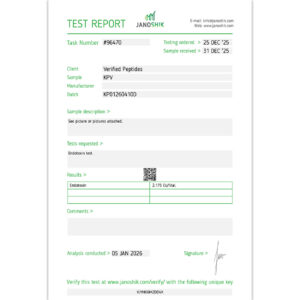
This batch of KPV has been third party lab tested and verified for quality.

TESTED FOR:

purity
weight
TFA FREE
sterility
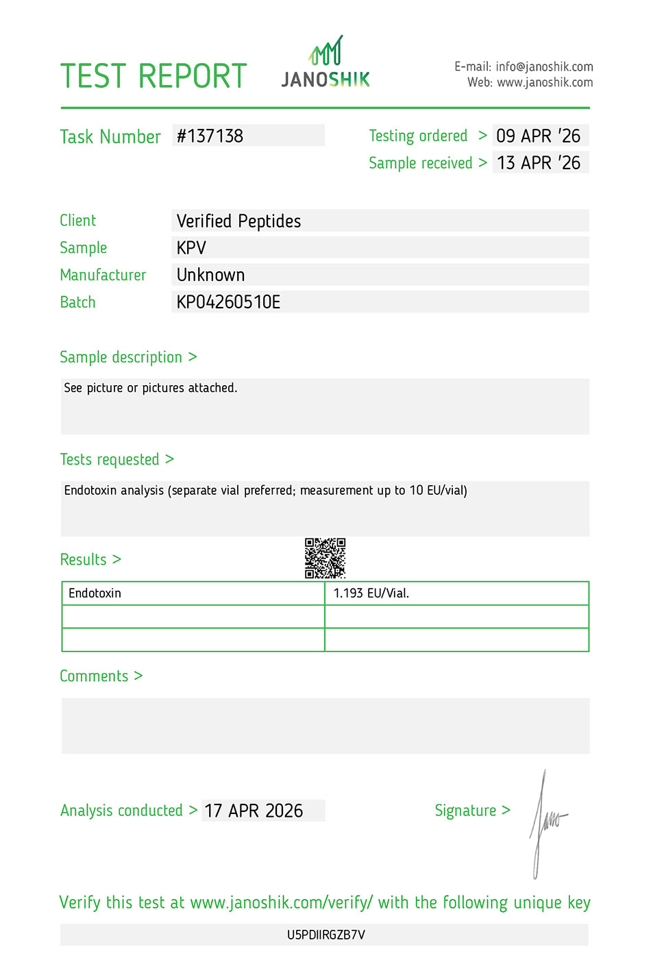
endotoxins(lps)
$55.00
| Quantity | Price | |
|---|---|---|
| 0% | 1 - 4 | $55.00 |
| 10% | 5 - 9 | $49.50 |
| 20% | 10 - 19 | $44.00 |
| 30% | 20 + | $38.50 |
This product comes as lyophilized powder and is not reconstituted. All products and materials sold on this site are for Research Use Only and not for human use; subject to our Terms and Conditions.

2-Day Shipping
Order by 2:00 PM ET

INSULATED SHIPPING
Ice foam-box shipping available
| Peptide vial specifications | Results | |
|---|---|---|
Not reconstituted | ||
Bottle type: | Vial - sealed - flip top | |
Form: | Powder (lyophilized) | |
Vial size: | 3ML | |
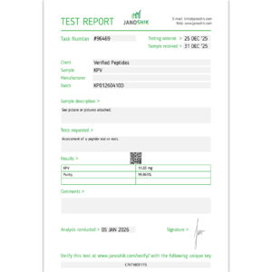
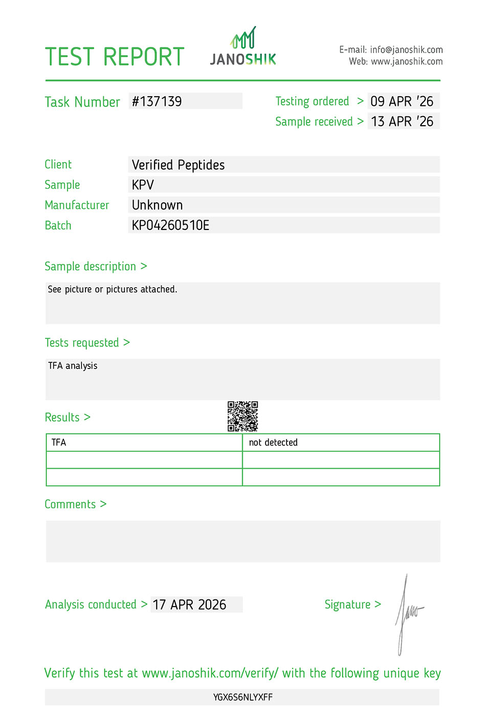
KPV Certificate of Analysis (COA) Test Result
| Date Tested: | |
|---|---|
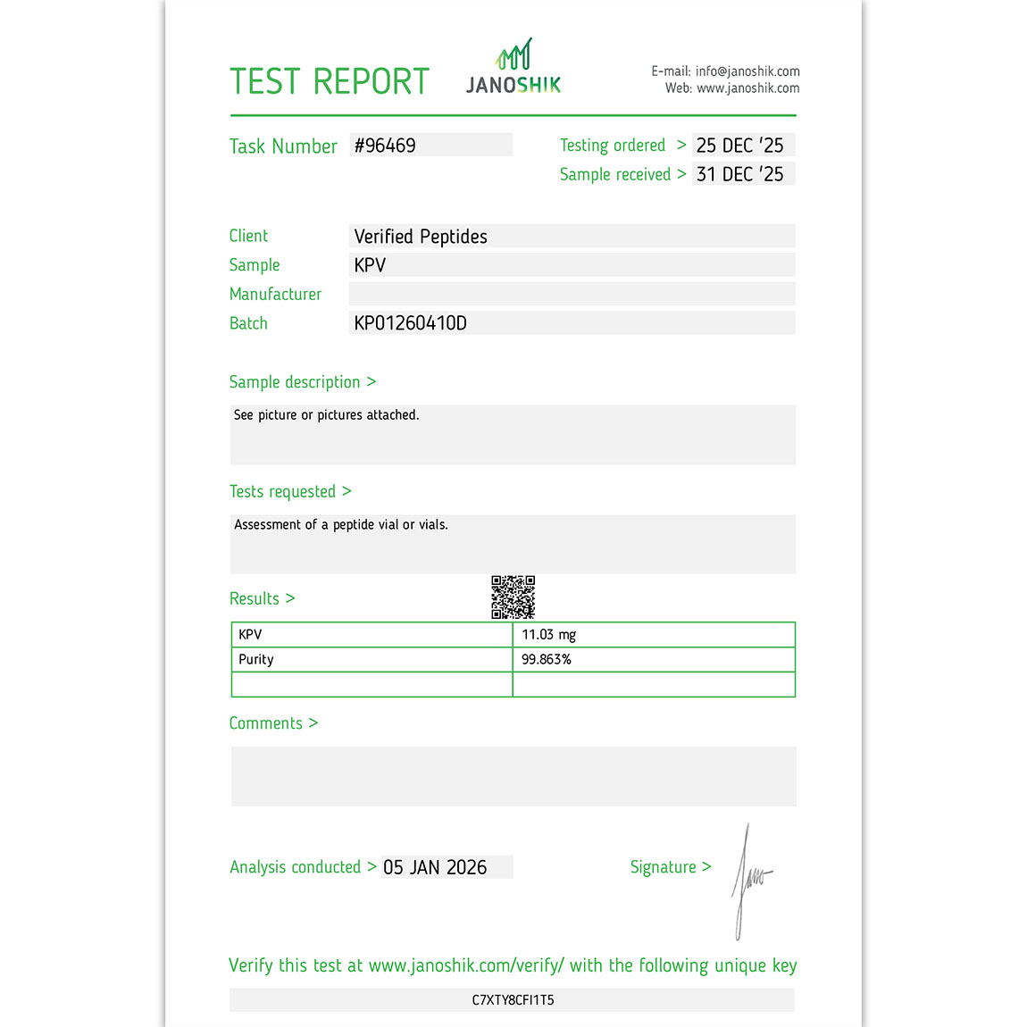
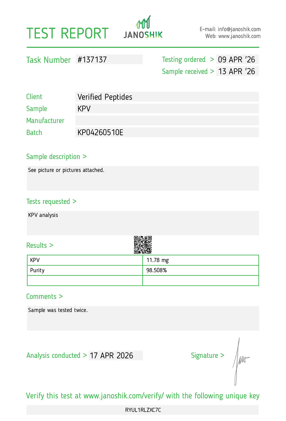
| Purity: | 99.508% |
| Weight: | 11.78mg |
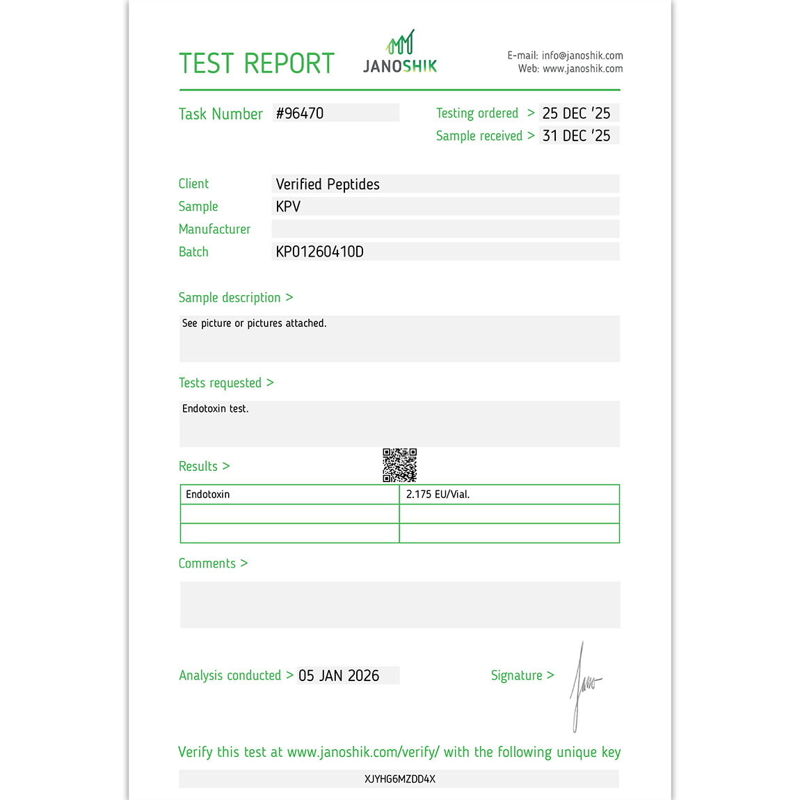
| Endotoxins (LPS): | Pass |
| Sterility: | Pass |
| TFA: | Pass |
| Batch #: | KP04260510E |
| Testing Lab: | Janoshik |
| COA Verified Link: | Purity Report Endotoxin Report |
| Testing Methods: | HPLC, LAL, TAMC/YAMC USP <71> |
KPV Specifications: Molecular Weight, CAS Number & Sequence
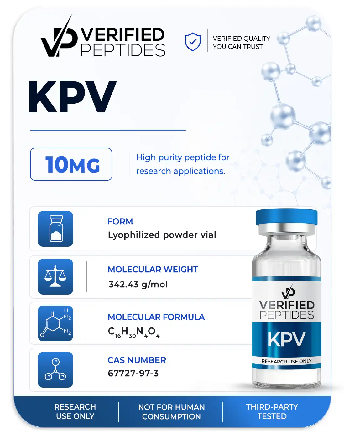
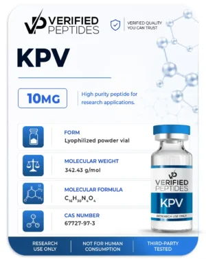
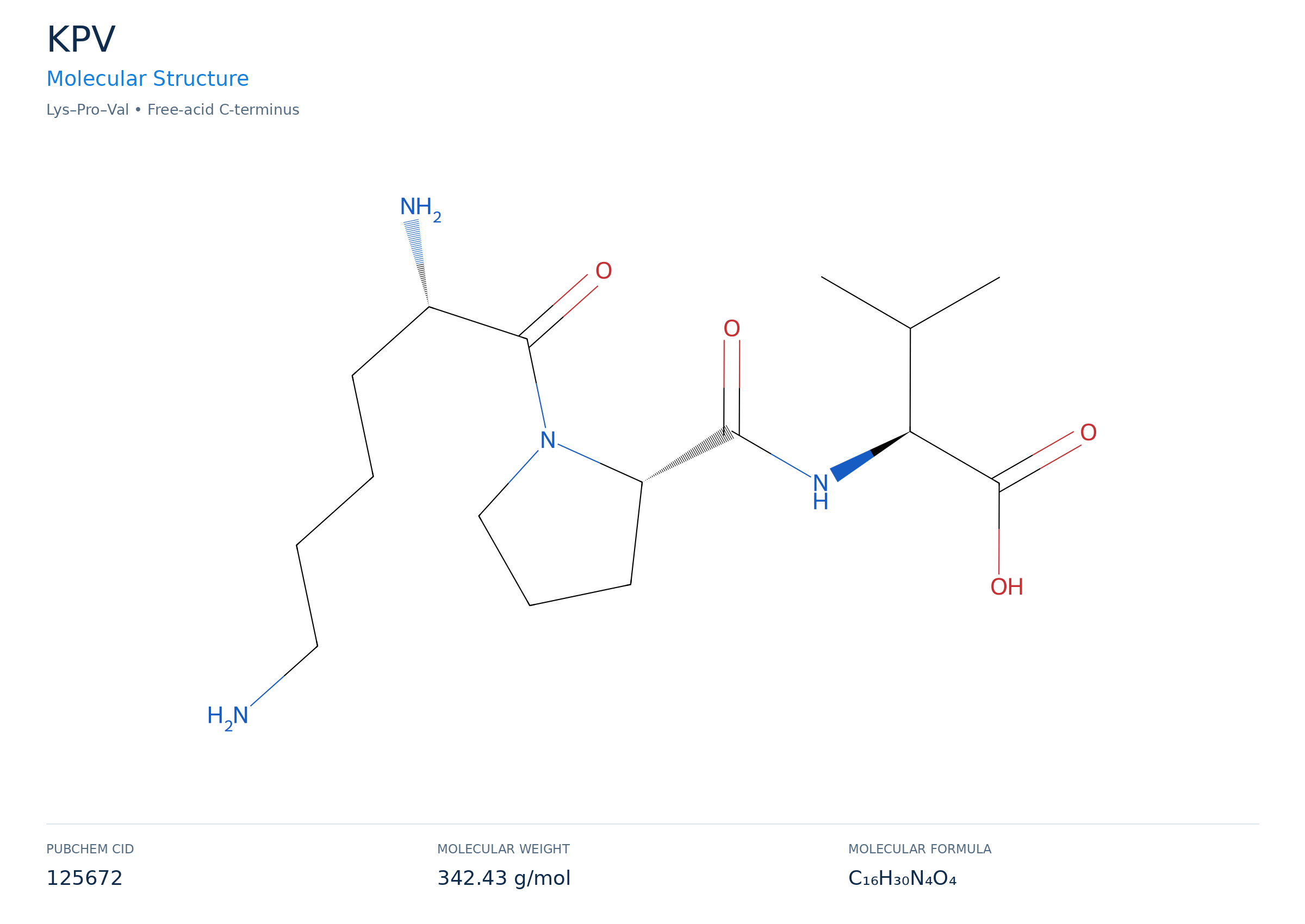
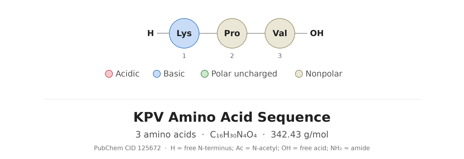
KPV (CAS 67727-97-3) is a tripeptide with the molecular formula C16H30N4O4 and a molecular weight of 342.43 g/mol.
| Form: | Powder (lyophilized) |
|---|---|
| Weight: | 10mg |
| Appearance: | White lyophilized powder |
| CAS Number: | 67727-97-3 |
| Molecular Formula: | C16H30N4O4 |
| Molecular Weight: | 342.43 g/mol |
| PubChem CID: | 125672 |
| Sequence: | Lys-Pro-Val |
| Other Names: | KPV, KPV Tripeptide, Lysine-Proline-Valine, α-MSH(11-13) |
| Terms: | Subject to our Terms and Conditions. This material is sold for laboratory research use only. Not for human consumption, animal, or medical use. |
KPV Molecular Structure
KPV Amino Acid Sequence

The industry's most trusted source for research peptides
The Leader In Peptide Testing
500+ Certificates of Analysis
You can be sure we don't cut corners on testing every single batch.
Visit our Lab Reports page.
An Established, Trusted Company
6+ Years of Peptides
We pioneered third party lab testing and have stood the test of time. Our first lab report dates back to 2019 proving our track record.
Fair market pricing
Competitively priced cost per milligram for verified quality.
Peptides Tested For More Than Purity
We conduct an array of analytical testing for our peptides:
purity, weight, endotoxins(LPS), sterility (bacteria & mold/yeast), and TFA content.
Transparent. Consistent. Reliable.
- Batch number on every vial
- Published Certificates of Analysis
- Expert customer service
- Satisfaction and quality guarantee
- Cold pack foam-lined shipping
Explore Other High-Purity Peptides
Peptide Blends
Price range: $65.00 through $107.00
Peptides
$49.99
Peptides
$44.99
Peptides
$62.00
Peptides
$119.00
Peptides
Price range: $43.00 through $77.00
Peptides
$34.99
Hear What Our Customers Say
Trusted by thousands of researchers for high-quality peptides

This is a first class operation. Quality products and the best customer service. There may be many similar type of companies out there, but this one is trusted and affordable. [...] I highly recommend this company.
Verified Peptides is hands down my favorite website for these products. I can always count on the products to be legitimate and effective, all while being affordable. Will continue to give them my business.
Outstanding product, outstanding price, outstanding shipping
What is KPV?
KPV is a tripeptide (lysine–proline–valine) that matches amino acids 11–13 of α-melanocyte-stimulating hormone (α-MSH). It is also written as α-MSH (11–13). α-MSH is a melanocortin peptide derived from the precursor protein pro-opiomelanocortin (POMC). Researchers use KPV in cell and animal studies to examine which parts of the α-MSH sequence are linked to its effects on inflammatory signaling.
KPV Peptide Research Studies
Summaries of peer-reviewed research. For research and educational purposes only.
🧫 Peer-Reviewed Study · In Vitro Cell Culture
KPV Cell Culture Study: Melanocortin Peptides and Cytokine Production in Mouse Microglia (Delgado et al., Journal of Leukocyte Biology, 1998)
Overview of the KPV Study
This 1998 study in the Journal of Leukocyte Biology (Delgado et al.) tested three melanocortin peptides in a mouse microglial cell line: full-length α-MSH (1–13), the tripeptide KPV, written as α-MSH (11–13), and ACTH (1–24). Microglia are the resident immune cells of the brain.
Cells were stimulated with bacterial lipopolysaccharide (LPS) plus interferon-γ (IFN-γ) to trigger an inflammatory response. Researchers then measured tumor necrosis factor-α (TNF-α), interleukin-6 (IL-6) and nitric oxide (NO). They also examined cAMP signaling, gene expression, and whether the microglia produce α-MSH themselves.
Scope: All experiments were performed in a cultured mouse cell line. The study did not include animals or people.
KPV Study at a Glance
- Citation: Delgado R, Carlin A, Airaghi L, et al. J Leukoc Biol. 1998;63(6):740–745.
- Study type: In vitro cell culture
- Cells: N9 immortalized mouse microglial cell line
- Stimulus: LPS (10 ng/mL) plus IFN-γ (1 U/mL) for 24 hours
- Peptides compared: α-MSH (1–13), α-MSH (11–13) (KPV), ACTH (1–24)
- Measurements: TNF-α, IL-6 and nitrite (a marker of NO), intracellular cAMP, TNF-α and iNOS mRNA, and α-MSH released by the cells
Why Researchers Study KPV and Melanocortin Signaling in Microglia
Activated microglia release cytokines and nitric oxide that help defend the brain against infection. In excess, these mediators are linked to tissue injury in experimental models. Earlier work had shown that α-MSH reduces inflammatory mediators in peripheral immune cells such as monocytes and macrophages, and that those cells can make α-MSH themselves.
This study asked three questions about microglia:
- Do melanocortin peptides, including the short KPV fragment, reduce TNF-α, IL-6 and NO production?
- Do microglia release α-MSH when activated?
- Does blocking that endogenous α-MSH change the inflammatory response?
Experimental Design and Methodology
N9 cells were plated and treated with each peptide 10 minutes before LPS plus IFN-γ. In pilot work, the peptides were tested from femtomolar to millimolar concentrations. The authors chose micromolar concentrations for the main experiments because they gave the most consistent inhibition under these culture conditions. Cell viability stayed above 98% in all conditions.
The study used several assays:
- TNF-α by cell bioassay, IL-6 by ELISA, and nitrite by the Griess reaction
- Intracellular cAMP after peptide exposure, with forskolin as a comparison
- Northern blots for TNF-α and inducible nitric oxide synthase (iNOS) mRNA
- α-MSH released into the medium, measured by radioimmunoassay
- Immunoneutralization of endogenous α-MSH with an anti-α-MSH antibody, compared with control IgG
What Did the KPV Study Find? Cytokine and Nitric Oxide Results
All three melanocortin peptides reduced TNF-α, IL-6 and nitrite production by activated microglia compared with stimulated cells without peptide:
- α-MSH (1–13): The greatest inhibition was at 10 µM, about 43% (TNF-α), 31% (IL-6) and 42% (nitrite). The concentration–response pattern was U-shaped, as earlier α-MSH studies had reported.
- KPV, α-MSH (11–13): About 45% (TNF-α), 50% (IL-6) and 40% (nitrite)
- ACTH (1–24): About 38% (TNF-α), 65% (IL-6) and 41% (nitrite)
The authors noted that the three-amino-acid KPV fragment was "likewise a very effective inhibitor" in this assay system.
Gene Expression and cAMP Signaling
Northern blot analysis showed that the lower TNF-α and nitrite output matched lower TNF-α mRNA and iNOS mRNA. This indicates the effect occurred at the level of gene expression.
Both α-MSH (1–13) and KPV increased intracellular cAMP in resting cells and in cells stimulated with LPS plus IFN-γ. The size of the increase was comparable to that caused by forskolin, an adenylyl cyclase activator. Citing earlier studies in which other cAMP-raising agents reduced TNF-α and iNOS in microglia, the authors proposed that increased cAMP probably contributes to the effects they observed.
Do Microglia Produce α-MSH?
Microglia stimulated with LPS plus IFN-γ released α-MSH into the culture medium. When endogenous α-MSH was neutralized with an antibody before and during stimulation, release of IL-6, TNF-α and nitrite rose by about 26%, 28% and 27% compared with cells given control IgG. α-MSH was undetectable in the antibody-treated samples.
The authors interpreted this as evidence of an autocrine circuit: activated microglia release α-MSH, which in turn limits their own production of inflammatory mediators in culture.
An Open Question: Which Receptor Does KPV Use?
Five melanocortin receptors (MC1R to MC5R) had been identified by 1998, and all signal through cAMP. At that time, none had been shown to bind KPV. Earlier work found that KPV did not compete with α-MSH for binding at MC1R on melanoma cells. The authors left open whether KPV acts through a known melanocortin receptor in an unusual way or through a different, uncharacterized receptor.
Limitations and Research Context
- All results come from one immortalized mouse microglial cell line. Primary microglia or cells from other species may respond differently.
- Culture conditions, including the strong LPS plus IFN-γ stimulus and micromolar peptide concentrations, are specific to this experiment.
- A cell culture cannot model interactions with neurons, astrocytes, blood vessels and other cells in living tissue.
- KPV's receptor and mechanism were not resolved in this study.
- In vitro results do not establish effects, safety or appropriate exposure in animals or people.
Conclusion
Delgado et al. (1998) found that α-MSH (1–13), KPV and ACTH (1–24) each reduced TNF-α, IL-6 and nitric oxide production by activated mouse microglia in culture. The effects were accompanied by lower TNF-α and iNOS mRNA and higher intracellular cAMP. The microglia also released α-MSH, and blocking it increased inflammatory output by roughly 26–28%, which suggests an autocrine regulatory loop in this cell model.
This summary is provided for research and educational purposes. Statements on this page have not been evaluated by the FDA. Research compounds offered on this site are for laboratory research use only. They are not intended for human or veterinary use, or to diagnose, treat, cure, mitigate or prevent any disease.
Frequently Asked Questions
What is KPV, and how is it related to α-MSH?
KPV is the tripeptide lysine–proline–valine, equal to amino acids 11–13 at the C-terminal end of α-melanocyte-stimulating hormone. It is often written as α-MSH (11–13).
What did the 1998 KPV microglia study find?
In mouse N9 microglia stimulated with LPS plus IFN-γ, KPV reduced TNF-α by about 45%, IL-6 by about 50% and nitrite by about 40%. Full-length α-MSH and ACTH (1–24) produced reductions of similar size.
How did the authors explain KPV's effect on cytokines in microglia?
KPV and α-MSH both raised intracellular cAMP to levels comparable to forskolin, and both lowered TNF-α and iNOS mRNA. Based on this and earlier studies, the authors proposed that cAMP signaling probably contributes to the reduced cytokine and NO production.
Do microglia make their own α-MSH?
In this study, activated microglia released α-MSH into the culture medium. Blocking it with an antibody raised IL-6, TNF-α and nitrite release by about 26%, 28% and 27%, which suggests an autocrine feedback loop in culture.
Which receptor does KPV act on?
The study did not resolve this. In 1998 none of the five known melanocortin receptors had been shown to bind KPV, and earlier work found that KPV did not compete with α-MSH at MC1R.
Study Reference
Delgado R, Carlin A, Airaghi L, Demitri MT, Meda L, Galimberti D, Baron P, Lipton JM, Catania A. Melanocortin peptides inhibit production of proinflammatory cytokines and nitric oxide by activated microglia. J Leukoc Biol. 1998;63(6):740–745. doi:10.1002/jlb.63.6.740
https://pubmed.ncbi.nlm.nih.gov/9620667/
IN THE KNOW
Join over 5,000 active members and get updates on new arrivals and latest lab reports.