
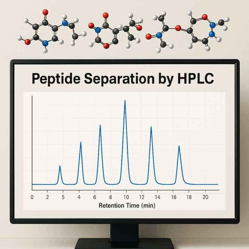
Peptide Separation by HPLC: Precision for Protein Research
Author: Dr. Numan S. Date: August 14, 2025
Why Peptide Separation by HPLC Remains a Gold Standard
High-performance liquid chromatography (HPLC) has been the workhorse for peptide analysis for decades. It earned its reputation by providing exceptional resolution and consistency in separating peptide mixtures. In fact, HPLC is widely regarded as the gold standard for isolating and purifying peptides [1]. The reason is its ability to handle complex peptide samples and yield sharp, well-resolved peaks, which is crucial for both research and quality control. This robust performance has revolutionized protein and peptide analysis, enabling scientists to separate peptides of varying sizes and properties with precision [2].
Another advantage is the method’s versatility. HPLC methods can be fine-tuned by adjusting solvents, gradients, and columns to suit different peptide characteristics. Compared to older techniques, HPLC offers superior sensitivity and scalability – from analytical microgram levels up to preparative scales for milligram quantities. The technique’s reliable retention time reproducibility means that peptide peaks can be identified consistently across runs, which is vital for protein analysis workflows like peptide mapping. All these factors explain why HPLC is used for peptide separation in laboratories worldwide: it delivers trustworthy, high-resolution separations that form the backbone of modern proteomics and peptide chemistry.
Types of HPLC Used in Peptide Analysis
Peptide scientists leverage several modes of HPLC, each exploiting different chemical properties of peptides to achieve separation [2]. The major types of HPLC used in peptide mapping and purification include:
- Reversed-phase HPLC (RP-HPLC): The most commonly used mode for peptides. It separates peptides based on hydrophobicity – peptides interact with a nonpolar stationary phase (often C18 silica), and a gradient elution of increasing organic solvent (e.g., acetonitrile) elutes peptides in order of increasing hydrophobicity. RP-HPLC offers high resolution and sensitivity, which is why it remains the preferred separation mode for peptides [3]. Peptides with more nonpolar amino acids have longer retention and elute later in the gradient. This method is excellent for peptide mixtures and is the default for HPLC peptide analysis in proteomics.
- Ion-exchange HPLC (IEX): This mode separates peptides by charge. Cation-exchange columns bind positively charged peptides, whereas anion-exchange columns bind negatively charged peptides. By gradually changing salt or pH, peptides elute based on their charge properties. Ion-exchange is especially useful as a complementary step – for example, enriching peptides by charge class before a final RP-HPLC run. It can provide better resolution for peptides that differ mainly in net charge (such as variants or modifications).
- Size-exclusion HPLC (SEC): Also known as gel filtration, SEC separates molecules by size. Smaller peptides take longer to elute because they penetrate the pores of the column packing, whereas larger peptides elute faster. SEC is used more for protein separations or to check aggregation states, but in peptide work it can confirm conjugate formation or aggregate removal. Its resolving power for peptides is lower than RP-HPLC, so it’s often reserved for assessing peptide purity and high-level size differences rather than fine analytical separations.
- Hydrophilic Interaction Liquid Chromatography (HILIC): HILIC is essentially the opposite of reversed-phase – it uses a polar stationary phase and an organic-rich mobile phase to retain polar peptides. Extremely hydrophilic or highly charged peptides that elute too early (or not at all) in RP-HPLC may be better separated by HILIC. This technique has gained traction for certain post-translationally modified peptides or glycopeptides that RP-HPLC struggles with.
- Mixed-Mode Chromatography: Emerging analytical chromatography media combine multiple interactions in one column. For instance, a mixed reversed-phase/ion-exchange column can separate peptides by both hydrophobicity and charge simultaneously. This approach, discussed more under trends, can resolve complex peptide mixtures that single-mode columns cannot. Mixed-mode columns are especially useful for challenging separations, such as very polar peptides or peptide variants, by providing complementary retention mechanisms in one step.
Each HPLC mode offers unique selectivity. Often, peptide separation by HPLC employs a combination of these techniques – for example, an ion-exchange step to group peptides by charge, followed by RP-HPLC to fine-tune separation by hydrophobicity. The choice of method depends on the nature of the peptides and the goals of the analysis.
Choosing the Right Column and Mobile Phase
Selecting an appropriate HPLC column and mobile phase is critical for efficient peptide separation. Column choice largely centers on the stationary phase chemistry and pore size. For reversed-phase HPLC, silica-based C18 columns with wide pore sizes (~300 Å) are standard for peptide mapping, as they accommodate the folded structures of larger peptides and proteins. A longer alkyl chain (C18 vs C4) generally provides stronger hydrophobic interactions, but shorter chain columns or those with polar embedded groups can be useful for very hydrophilic peptides. Researchers often screen multiple columns because each can have slightly different selectivity. In fact, one study characterized dozens of RP-HPLC columns and created a database to help identify alternatives, highlighting how column selection impacts peptide peak resolution and peptide purity assessments. Matching the column’s properties to the peptide mixture (considering peptide size, hydrophobicity, and modifications) is a form of method optimization.
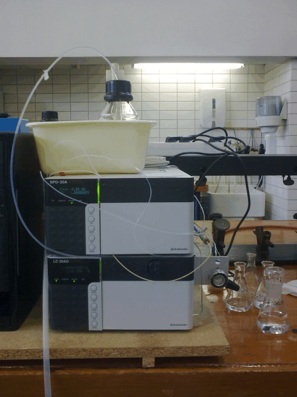
Figure 1: A typical HPLC system used for peptide separation by HPLC in proteomics and pharma labs.
Equally important is mobile phase optimization. Reversed-phase separations of peptides typically use a binary mobile phase: aqueous buffer with 0.1% acid (for example, trifluoroacetic acid, TFA) and an organic modifier (such as acetonitrile or methanol with the same acid). The acid in the mobile phase serves multiple roles. Traditionally, 0.1% TFA has been used because it acts as an ion-pairing agent and suppresses silanol interactions on the silica, yielding sharp peaks and good retention for peptides [3]. This is optimal for UV-based analytical detection and contributes to high separation efficiency. However, TFA in the mobile phase can drastically reduce sensitivity when using mass spectrometry detection, because it ion-pairs with peptides and suppresses their ionization. To address this, labs focusing on LC–MS integration often switch to weaker acids like formic acid or use lower TFA concentrations. Formic acid (around 0.1% or with ammonium formate buffer) still provides the necessary low pH but causes less ion suppression in the MS. The trade-off is that peptides might exhibit more peak tailing or slightly lower resolution with formic acid alone. To mitigate this, newer analytical chromatography columns have surface chemistries designed for formic acid compatibility, or laboratories may use additives like acetic acid or specialized ion-pairing agents (e.g. difluoroacetic acid) that balance chromatographic performance and MS sensitivity.
Ultimately, the “best” mobile phase for peptide separation depends on the detection method and peptide properties. If maximum resolution and peptide mapping detail are needed (for example, mapping a monoclonal antibody digest by UV), TFA with a slow gradient is often preferred for its unbeatable peak shape. If the goal is to identify peptides by MS, a formic acid-based mobile phase is usually recommended to preserve signal intensity, accepting a minor compromise in chromatographic sharpness. Gradient elution profiles also need optimization – a shallow gradient (slow increase in organic) can better resolve similar peptides, whereas a steeper gradient runs faster but might co-elute components. Method development often involves trying different gradient slopes, temperatures, and additives to achieve optimal separation. Comprehensive studies have shown that exploring a wide range of mobile-phase pH, additives, and organic solvents can maximize peptide chromatographic selectivity. In practice, finding the right column and mobile phase is an iterative process of fine-tuning to balance resolution, run time, and compatibility with detection (UV or MS).
HPLC vs. Other Peptide Separation Techniques
Given HPLC’s dominance, it’s worth comparing it to alternative peptide separation techniques. Capillary electrophoresis (CE) is one such technique that analysts use for peptides. In CE, peptides migrate in an electric field inside a capillary, separating based on charge-to-size ratio rather than on a stationary phase. CE can achieve extremely high efficiency and has minimal sample requirement, making it a powerful complementary method for peptide analysis [2]. In fact, some protocols use CE alongside HPLC to gain additional separation of peptide variants. However, CE equipment and method development are less common in many labs, and the technique can be sensitive to matrix conditions. HPLC, in contrast, is widely available and easily coupled to detectors like UV and MS, which is a major reason it remains more popular for routine HPLC peptide analysis. The value of CE is often in niche applications or when HPLC methods aren’t resolving certain analytes – for example, very small peptides or highly charged ones might be better handled by CE, but overall HPLC’s robustness and integration with standard lab workflows give it an edge [2].
Another alternative is gel electrophoresis (e.g., SDS-PAGE), which separates proteins (and to some extent large peptides) by size in a polymer gel. While gels are indispensable for protein analysis, they have limited resolution for peptide mixtures and are not quantitative in the way HPLC is. Analytical chromatography techniques like HPLC also surpass gels in allowing direct coupling to mass spectrometry for peptide identification.
Emerging non-chromatographic methods are also being explored. For instance, peptide separation using magnetic nanoparticles or membrane-based methods can achieve purification without the high solvent consumption of HPLC. These novel techniques aim to rival HPLC’s performance with potentially lower cost or faster throughput. In recent studies, membrane filters and isoelectric focusing devices have shown ability to separate peptides with high speed, and researchers have demonstrated that some newer approaches can approach the resolution of established HPLC methods. However, these are still evolving, and they often complement rather than fully replace HPLC. For now, HPLC remains the gold standard for peptide separation in both research and industry, with other techniques serving as supplementary tools. The choice often comes down to the specific context: for regulated environments requiring validated methods and peptide purity data (such as pharmaceutical quality control), HPLC is almost always the technique of choice. In more exploratory research settings, a combination of HPLC and other methods (like CE or novel nanomaterial-based separations) can be employed to tackle particularly challenging peptide mixtures.
Common Challenges in Peptide Separation and How to Solve Them
Separating peptides is not without challenges. One common issue is dealing with peptides at the extremes of polarity. Very hydrophilic peptides (for example, ones rich in acidic or polar residues) may elute near the void volume in reversed-phase runs, with poor retention and resolution. On the other hand, extremely hydrophobic peptides might stick strongly and elute only at very high organic content or even precipitate. The limitation of HPLC in peptide analysis here is that a single mode may not adequately separate all components if their chemistries diverge greatly. A practical solution for polar peptides is to switch to a different mode like HILIC. As mentioned, HILIC provides better retention of polar species: using a polar stationary phase and high organic solvent can make those early-eluting peptides separate with decent resolution [2]. Conversely, for extremely hydrophobic or aggregated peptides, adding organic co-solvents (e.g. isopropanol) or using a shallow gradient and higher temperature can improve peak shape. Some peptides that tend to aggregate can be helped by including chaotropic agents or using smaller injection amounts to avoid column overload.
Another challenge is peak tailing and broadening, often observed for basic peptides on silica-based columns. This is caused by unwanted ionic interactions with residual silanol groups on the stationary phase. Tailing peaks not only reduce resolution but also complicate quantification. The remedy has been to use ion-pairing additives (like TFA) to neutralize silanols [4], or newer column technologies that “shield” or deactivate silanol sites. Modern RP columns often are end-capped or use surface modifications to minimize these interactions, resulting in more symmetric peaks even for peptides with multiple basic residues. Additionally, using an appropriate pH (most peptide HPLC is done at pH ~2–3) ensures amino groups are protonated and less apt to stick to silica.
Co-elution of peptides is another frequent hurdle, especially in complex mixtures like a tryptic digest of a protein. When dozens of peptides elute in a narrow timeframe, some peaks overlap, risking that important components go undetected. To solve this, analysts turn to method adjustments – for instance, lengthening the gradient (e.g., extending a 30-minute run to 60 minutes to spread peaks out) or employing multi-dimensional separation. An example of the latter is off-line fractionation: one can first separate peptides by ion exchange or by high-pH RP into fractions, then run each fraction by conventional low-pH RP-HPLC. This two-dimensional approach greatly increases overall peak capacity and can resolve peptides that co-eluted in a single run. While more labor-intensive, it’s a proven strategy in proteomics to deal with extremely complex samples.
Instrumental and practical issues can also arise. Carryover of peptides – when a bit of a peptide from a previous run sticks in the system and appears in the next run – can confuse results. This is mitigated by thorough column washing and using guard columns or passivating any metal surfaces in the flow path (peptides can adhere to metal tubing or injector parts). Another challenge is peptide stability: some peptides might degrade or stick to vial surfaces. Using silanized vials or adding mild stabilizers can preserve sample integrity.
Crucially, most challenges in peptide HPLC can be addressed through methodical optimization. Small changes in the mobile phase (such as adding an ion-pair agent, or a salt, or changing pH), adjusting column temperature, or trying a different stationary phase often lead to significant improvements in separation. Systematic method development – varying one factor at a time or using design-of-experiment approaches – is commonly employed to troubleshoot difficult separations. For example, if two peptides consistently co-elute, one might try mobile phase optimization by changing the slope of the gradient or using a different organic solvent (some peptides resolve better in methanol vs acetonitrile). As one primer on peptide isolation notes, factors like solvent choice, modifier, gradient, and temperature all impact the result, and method adjustments can enhance purity and yield when standard conditions fall short [7]. Ultimately, a deep understanding of peptide chemistry and chromatographic principles, combined with careful experimentation, allows scientists to overcome these challenges and achieve reliable separation – even for tricky peptide samples.
Applications in Drug Development and Proteomics
Peptide HPLC is central to both pharmaceutical development and proteomic research. In drug development, many therapeutic agents are peptides (or peptide-based conjugates), and ensuring these products are pure and correctly synthesized is paramount. Peptide purity analysis by HPLC is a routine quality control step for any synthesized peptide. Analytical RP-HPLC, often coupled with a UV detector at 214 nm (where peptide bonds absorb), is used to quantify the purity of a peptide batch by separating impurities or deletion sequences from the main product. Peptide manufacturers typically require a purity of >95% for clinical-grade peptides, and HPLC provides the quantitative readout to support that. For instance, if you order a synthetic peptide, the certificate of analysis will usually include an HPLC chromatogram showing a dominant main peak and any minor impurity peaks with their percentage areas. Peptide separation by HPLC thus directly informs whether a peptide is suitable for therapeutic use or needs re-purification. In fact, regulatory guidelines call for impurity profiling of peptide drugs, and HPLC (often RP-HPLC combined with mass spectrometry for identity confirmation) is the endorsed method to perform this. Some specialized HPLC methods (such as ion-exchange HPLC) are also used to characterize variants of synthetic peptides – for example, to ensure there are no charge-altered variants or to quantify deamidation levels in a peptide drug.
Beyond purity, HPLC helps in characterizing peptide modifications. Many peptide drugs are modified (pegylated, lipidated, etc.) or have disulfide bonds. Reversed-phase HPLC will often be used to verify that the modification is present (since it usually shifts the peptide’s retention time). In conjugated peptides, a separate peak might correspond to the unconjugated form, which HPLC can quantify. Thus, HPLC supports both the purification process and the final quality assessment of peptide therapeutics.
In proteomics and protein analysis, HPLC is indispensable in the form of LC–MS/MS workflows. Here, complex protein mixtures (like cell lysates) are enzymatically digested into peptides, which are then separated by HPLC before entering a mass spectrometer for identification. This LC-MS integration marries the separation power of HPLC with the analytical power of MS. The result has been a revolution in proteomics: LC–MS is now considered the gold standard methodology for identifying and quantifying proteins in complex samples. The HPLC step is critical because the mass spectrometer performs best if peptides enter one at a time; a good chromatographic separation increases the number of distinct peptides that can be detected and quantified in a single run. Typically, nano-scale reversed-phase HPLC is used for proteomic peptide mapping, using long, very small diameter columns to achieve high resolution with very low flow rates compatible with electrospray ionization. The term peptide mapping in a proteomic context refers to the pattern of peptide peaks that represent a protein’s fingerprint. By comparing peptide maps, researchers can confirm a protein’s identity (each protein produces a characteristic set of peptides and thus chromatographic peaks) and even locate post-translational modifications (which often cause shifts in retention or the appearance of additional peaks). For example, a deamidation or oxidation in a peptide might produce a slightly different retention time or an extra peak, alerting analysts to that modification.
HPLC coupled to MS (LC–MS/MS) in proteomics not only identifies peptides but also allows label-free or isotope-labeled quantification of proteins across samples. The reproducibility of retention times and peak areas in HPLC underpins the quantitation: peptides serve as proxies for their parent proteins, and consistent chromatography ensures that the same peptide is measured in each sample run. The development of high-throughput proteomics has in large part depended on improvements in HPLC (like autosamplers, nano-flow pumps, and column technologies) to handle thousands of peptides in a single analysis.
In summary, whether it’s confirming the peptide purity of a new drug candidate or mapping the proteome of a cell, HPLC plays a pivotal role. It provides the accuracy, precision, and compatibility with detection methods required in these applications. It’s no exaggeration that without HPLC’s contributions to peptide separation, the fields of biopharmaceutical development and modern proteomics would not be as advanced as they are today.
Emerging Trends in HPLC for Peptide Mapping
The landscape of HPLC technology is continually evolving, and several emerging trends are enhancing peptide mapping and analysis capabilities. One major trend is the move toward ultra-high-performance liquid chromatography (UHPLC) systems. UHPLC uses columns packed with sub-2 μm particles and higher-pressure pumps. This allows much faster separations and improved resolution compared to conventional HPLC. For peptide separations, UHPLC can sharpen peaks and shorten analysis times, which is advantageous when dealing with large sample sets or when very high resolution is needed.
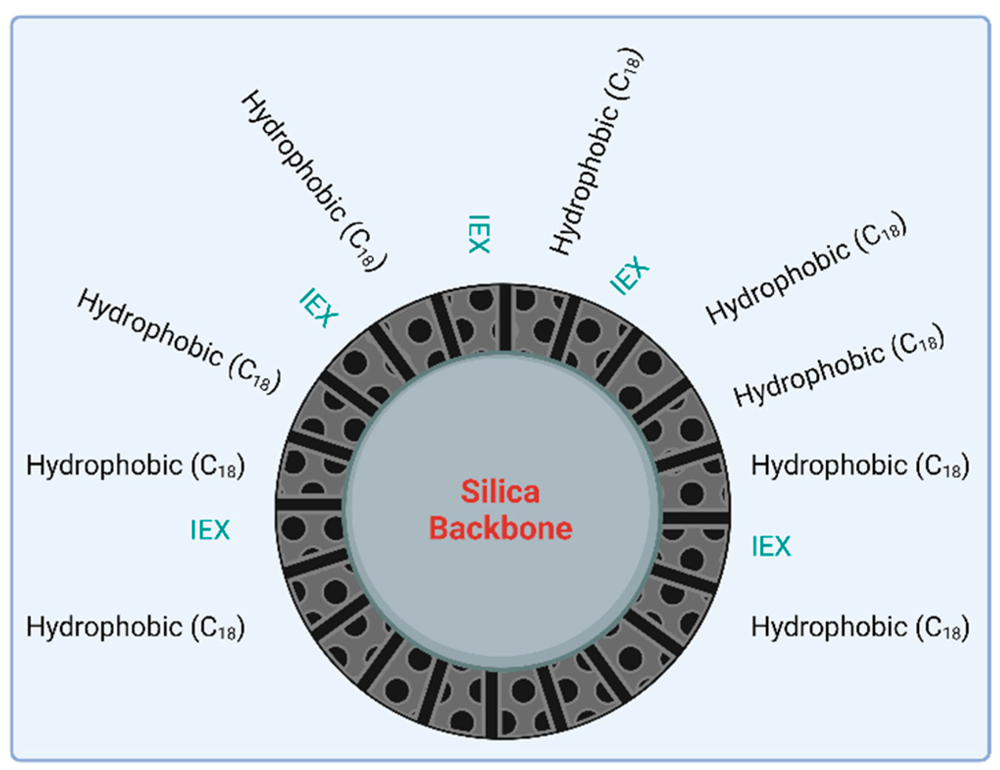
Figure 2: Schematic of a modern mixed-mode HPLC stationary phase combining hydrophobic C18 ligands (black) and ion-exchange (IEX) groups (teal) on the same silica support.
For example, a peptide map that might take 2–3 hours by traditional HPLC could potentially be run in under 1 hour by UHPLC without losing information. The trade-off historically was that UHPLC could be less forgiving (e.g., higher backpressure, more sensitivity to matrix) and sometimes slightly less reproducible, but improvements in instrument design have mitigated these issues. Many labs are now adopting UHPLC (or fast LC) for peptide analysis to increase throughput. Instrument vendors have even introduced systems that can handle both HPLC and UHPLC methods on the same platform, easing the transition for regulated labs. We also see gradient elution techniques being optimized for UHPLC – for instance, using segmented gradients or rapid ramping to make sure even very hydrophobic peptides elute within shorter run times.
Another trend is the development of new stationary phase chemistries and column formats. Analysts are experimenting with alternate bonded phases (beyond the standard C18). Examples include phenyl phases, pentafluorophenyl (PFP) phases, and mixed-mode phases, which can offer different selectivity. Notably, mixed-mode chromatography has gained interest for peptide mapping. These columns incorporate multiple interaction types, such as hydrophobic and ionic, in one column. The result is often a better separation of peptides that are hard to distinguish by hydrophobicity alone.
By using mixed-mode or multimodal columns, researchers have reported improved resolution of peptide variants and detection of minor species that might co-elute on conventional columns. This is especially useful in peptide mapping of protein therapeutics, where one needs to separate nearly identical peptides (e.g., oxidized vs. reduced forms, deamidated vs. native peptides). We can expect to see more of these innovative column types in mainstream use as they become commercially available.
Automation and integration are also key trends. Mobile phase optimization and method development are being accelerated through software and AI-driven tools. Automated method development systems can run many slight variations of a method (different pH, different gradient shapes) overnight and then suggest the best conditions, saving considerable time. In peptide mapping, where a method might need to be tailored for each new protein or product, such automation can greatly enhance productivity.
The integration of HPLC with detection is also advancing. While UV and MS are standard detectors for peptides, there’s growing interest in adding detectors like fluorescence (for naturally fluorescent tags), charged aerosol detectors, or even compact mass detectors to HPLC systems. For instance, a peptide mapping workflow might use UV absorbance for quantification and a low-cost mass detector for quick confirmation of peak identity, without needing a full high-end MS/MS on every run. This kind of multi-detector setup can improve the information obtained from each HPLC run.
Lastly, sustainability and green chemistry considerations are influencing HPLC practices. Traditional peptide HPLC uses a lot of acetonitrile and other solvents. Emerging trends include exploring solvent recyclers, smaller-volume columns (nano and micro flow HPLC use far less solvent), and even supercritical fluid chromatography for certain peptide separations to reduce solvent usage. While the core principles of peptide HPLC remain, these incremental innovations are making the technique faster, more powerful, and more adaptable to future needs.
In conclusion, peptide separation by HPLC continues to be a cornerstone of protein research, and ongoing improvements in technology ensure it will remain so. Whether through UHPLC’s speed, novel mixed-mode columns’ selectivity, or better integration with mass spectrometry, the precision and reliability of HPLC in HPLC peptide analysis only continue to grow. These advancements promise even more accurate and high-throughput peptide mapping – benefiting everything from academic proteomics studies to the development of the next generation of peptide therapeutics.
Frequently asked questions (FAQs) about Peptide Separation
What is the most effective method for separating peptides in solution?
- The most effective method for separating peptides in solution is typically High-Performance Liquid Chromatography (HPLC), particularly reversed-phase HPLC. This technique separates peptides based on their hydrophobicity and size, allowing researchers to achieve high purity in peptide samples. Other methods, like ion-exchange chromatography or size-exclusion chromatography, may also be used depending on the specific properties of the peptides being studied.
How does reversed-phase HPLC enhance peptide resolution?
- Reversed-phase HPLC enhances peptide resolution by using a hydrophobic stationary phase that interacts with the hydrophobic regions of peptides. This results in differential retention times for peptides with varying degrees of hydrophobicity. The separation is achieved by applying a gradient of solvents that vary in polarity, allowing peptides to elute at different times. The enhanced resolution makes it easier to isolate and identify individual peptides in a complex mixture.
What parameters impact peptide retention time in HPLC?
Several parameters impact peptide retention time in HPLC, including:
- Solvent composition: The polarity of the mobile phase affects how peptides interact with the stationary phase.
- Column type: Different columns with varying stationary phase characteristics can influence peptide interaction and retention.
- Flow rate: Higher flow rates typically reduce retention time, while lower flow rates increase it.
- Temperature: Changes in temperature can affect peptide interactions with the column and, subsequently, their retention times.
- Peptide properties: The amino acid sequence, size, and charge of the peptide all play significant roles in its retention during the chromatography process.
Why is peptide purity critical in drug research?
- Peptide purity is crucial in drug research because impurities can affect the biological activity, stability, and safety of peptide-based drugs. Inconsistent purity levels can lead to variability in experimental results, making it difficult to assess a peptide’s efficacy or potential side effects. Moreover, impurities may alter the mechanism of action or introduce harmful substances, making it essential to ensure high purity levels in peptides used for therapeutic purposes.
How is HPLC integrated with mass spectrometry in proteomics?
- HPLC is commonly integrated with mass spectrometry (MS) in proteomics to provide both high separation power and precise molecular identification. In this setup, peptides are first separated by HPLC based on their physical and chemical properties. The eluted peptides are then introduced into the mass spectrometer, where they are ionized and analyzed based on their mass-to-charge ratio. This integration enables researchers to identify and quantify peptides in complex biological samples, such as proteins, and provides detailed insights into protein structure, function, and interactions.
References
- Al Musaimi O, Jaradat DMM. Advances in Therapeutic Peptides Separation and Purification. Separations (MDPI). 2024;11(8):233. DOI:10.3390/separations11080233 mdpi.com
- Mant CT, Hodges RS. HPLC Analysis and Purification of Peptides. In: Methods in Molecular Biology, vol. 386: Peptide Characterization and Application Protocols. Humana Press; 2007:3-26. scispace.com
- Sigma-Aldrich (Merck KGaA). Eliminate TFA and Improve Sensitivity of Peptide Analyses by LC/MS. Supelco Application Note 168; 2002. sigmaaldrich.com
- Blackwell A. UV, MS, TFA, and Formic Acid – What to use? Peptide Mapping Part II. Agilent Community Blog. 2019. community.agilent.com
- JPT Peptide Technologies. Peptide Quality & Purity – Peptide Purity is routinely determined by HPLC. JPT Technical Resources; 2021. jpt.com
- Meston D, Stoll DR. Pitfalls in Proteomics: Avoiding Problems Before Data Acquisition. LCGC Europe. 2022;35(10):450-453. chromatographyonline.com
- Waters Corp. Practical Approaches to Peptide Isolation: Method Development Considerations. Waters Primer Application Note; 2018. waters.com