
Science Behind Electrospray Ionization in Peptide Analysis
Author: Dr. Numan S. Date: July 5, 2025
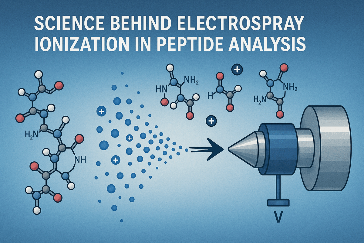
How a Mist of Charged Droplets Changed Peptide Science Forever
Mass spectrometers historically faced a strict upper mass limit for analytes – anything much above 1000 Da was nearly impossible to vaporize and ionize intact. This all changed in the late 1980s when electrospray ionization (ESI) was introduced (alongside MALDI, another “soft” ionization), effectively removing the molecular weight restriction and allowing even large peptides and proteins to be ionized and detected. ESI made it possible to “make elephants fly” – in other words, to transfer biomolecules of tens of thousands of daltons into the gas phase as ions without breaking them apart [5]. The development was so revolutionary that John B. Fenn, who pioneered ESI for biomolecules, shared the 2002 Nobel Prize in Chemistry for this advance.
ESI’s impact on peptide analysis and proteomics cannot be overstated. By producing a fine mist of charged droplets from a peptide solution, ESI enabled high-efficiency ionization of peptides at trace levels, which profoundly changed biomolecular sciences – now proteins and peptides could be identified and quantified from minute samples in high throughput. Peptide mixtures that once required extensive derivatization or were incompatible with older ionization techniques became accessible to mass spectrometry. In fact, electrospray ionization mass spectrometry (ESI-MS) rapidly became a cornerstone of proteomics workflows, allowing complex peptide digests to be analyzed directly by LC-MS/MS with femtomole-level sensitivity.
Breaking It Down: How Electrospray Ionization Works
To understand ESI, it helps to break down the mechanism of electrospray ionization into its fundamental steps. At its core, ESI uses a strong electric field to create a fine spray of charged droplets from a liquid sample. A peptide-containing solution is pumped through a narrow metal capillary held at a high voltage (typically 2–5 kV). The electric field causes the emerging liquid at the capillary tip to form a Taylor cone, from which a micro-jet of liquid ejects and breaks into an aerosol of tiny, charged droplets [7]. This process occurs at atmospheric pressure and can be operated in positive or negative polarity depending on whether one wants to produce cations (protonated peptides) or anions. Because ESI creates ions directly from solution and at ambient pressure, it is readily interfaced with liquid chromatographic separation and does not require heating the sample – ideal for fragile, nonvolatile peptide molecules.
As these initial droplets travel in the surrounding bath gas (often aided by a nebulizer gas and heat), they undergo solvent evaporation. Evaporation causes the droplet to shrink, which in turn brings the like charges on its surface closer together. There is a limit to how much charge a droplet can hold for a given size (the Rayleigh limit). When a droplet has lost enough solvent that electrostatic repulsion overcomes surface tension, it undergoes a Coulomb fission (explosion), flinging apart into many smaller droplets. Each progeny droplet carries away a fraction of the charge from the parent. This evaporation and fission cycle may repeat through multiple generations of ever-smaller, highly charged droplets. The mechanism by which final gas-phase peptide ions emerge from these droplets is explained by two main theoretical models: the Ion Evaporation Model (IEM) and the Charge Residue Model (CRM).
What Makes ESI Ideal for Peptide Analysis?
Several characteristics make electrospray ionization especially well-suited for peptide analysis compared to other ionization techniques. First, ESI is a soft ionization method that preserves the integrity of peptide molecules. Little to no fragmentation occurs during the ionization step, so the molecular ion (or more accurately, a protonated pseudomolecular ion) of the peptide is almost always observed. This means the peptide structure remains intact through ionization, which is crucial for accurate mass determination and for subsequently performing structural analysis via tandem mass spectrometry if needed. The ions produced by ESI are also in a low-energy, stable state (not highly excited), so they are less prone to decomposing on their own. This gentleness helps in preserving non-covalent interactions and higher-order structures if one is analyzing protein–peptide complexes or other labile assemblies.
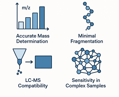
Figure 1: General workflow for isolating peptides from a biological sample.
From Sample to Signal: A Step-by-Step Look at the ESI Process
Illustration of the electrospray ionization process in positive-ion mode. (1) Sample introduction: A peptide-containing liquid (often a water/organic solvent mix with a little acid) is pumped through a fine needle held at high voltage. The strong electric field at the tip causes the liquid to form a Taylor cone and emit a spray of tiny, charged droplets (each droplet carrying excess positive charges in positive ESI) [7]. A mist of these charged droplets emerges from the electrospray needle. The formation of charged microdroplets constitutes the initial sample ionization step – the peptides are not yet free ions, but they are solvated in droplets that are electrically charged.
(2) Droplet evolution and disintegration: As the charged droplets travel away from the needle, a flow of heated nitrogen drying gas helps them evaporate solvent [7].The droplets rapidly shrink in size due to solvent evaporation, which increases the charge density on each droplet. Eventually, a point is reached (the Rayleigh limit) where the electrostatic repulsion between like charges on the droplet’s surface overcomes the surface tension holding the droplet together. At this moment the droplet undergoes a Coulomb explosion, ejecting smaller offspring droplets in an effort to shed charge and reach stabilityEach “fission” event releases a burst of smaller, more highly charged droplets. Importantly, throughout this evaporation and fission sequence, peptide molecules (and other solutes) become concentrated in the remaining droplet residue. Repeated cycles of evaporation and Coulomb fission continue until the droplets are extremely tiny – on the order of nanometers across. During these final stages, a droplet may contain only one peptide molecule (along with some solvent and charges). When that last bit of solvent evaporates, the peptide is left as a free gas-phase ion carrying multiple charges.
(3) Ion transfer and detection: The newly liberated peptide ions (e.g. [M+H]^+, [M+2H]^2+, [M+3H]^3+, etc.) are drawn into the mass spectrometer through a sampling orifice or capillary. This inlet typically has a slight vacuum and possibly an applied voltage to help attract the ions. Once inside the mass spectrometer’s high vacuum regions, the peptide ions are guided by ion optics into the mass analyzer (which could be a time-of-flight tube, an ion trap, a quadrupole filter, etc.). The analyzer separates ions by their mass-to-charge ratio, and a detector converts the arriving ions into electrical signals – a process of signal detection that ultimately yields the mass spectrum. The spectrum from ESI will often show a series of peaks corresponding to the same peptide with different charge states (for example, a peptide might show peaks at m/z values corresponding to [M+3H]^3+, [M+2H]^2+, [M+H]^+).
Single vs. Multiple Charging: Why It Matters in Peptide Workflows
One hallmark of electrospray ionization is that it produces multiply charged ions of peptides, whereas many other ionization techniques (notably MALDI) predominantly yield singly charged ions. In MALDI (Matrix-Assisted Laser Desorption/Ionization), a peptide typically appears as a single protonated ion [M+H]^+ in the spectrum. By contrast, in ESI a peptide commonly picks up multiple protons, resulting in ions like [M+2H]^2+, [M+3H]^3+, etc. [2]. The presence of these multiple charged ions means that a single peptide will produce several peaks (one for each charge state) in the mass spectrum – forming the characteristic charge state envelope. While this multiple charging adds complexity to the spectrum, it is extremely useful. The generation of multiple charges essentially divides the peptide’s mass by an integer number, so even very large peptides/proteins appear at a lower m/z. For example, a 30 kDa protein might produce a 30+ charge ion around m/z 1000, which is easily within the range of most mass analyzers. Multiply charged ions therefore extend the mass range of a given mass spectrometer, allowing analysis of high-mass biomolecules that would otherwise be beyond detection. This was one of the key reasons ESI opened the door to protein analysis in mass spectrometry – by yielding multiple charged ions, ESI-MS could “see” proteins that a singly-charged-ion technique would miss.
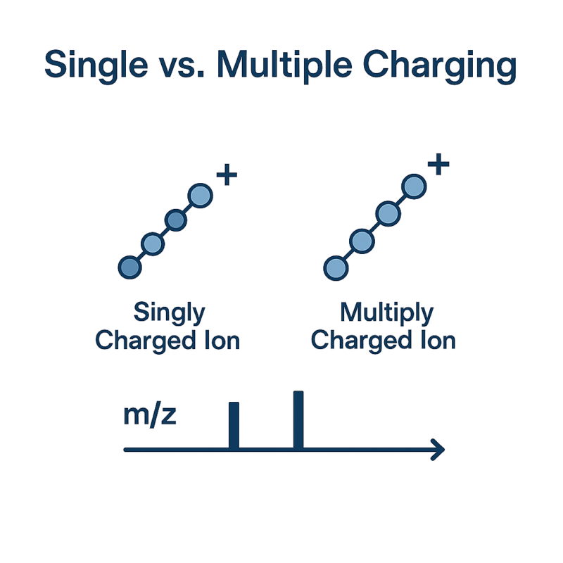
Figure 2: Difference between Single and Multiple Charging
Comparing ESI with Other Ionization Methods in Mass Spectrometry
ESI is one of several ionization techniques available in mass spectrometry, each with its pros and cons. Two of the most commonly used ionization techniques for biomolecules are ESI and MALDI, and they are often considered complementary. Both are soft ionization methods (minimal fragmentation) suitable for peptides and proteins, but they differ in how ions are generated. Electrospray ionization vs. MALDI: ESI generates ions from a liquid stream (continuous infusion or LC eluent) and produces multiple charged ions, whereas MALDI generates ions by laser desorption from a dried, solid matrix and predominantly yields single-charged ions [5].This fundamental difference leads to practical distinctions. MALDI’s singly charged ions make its spectra simpler (each peptide usually gives one peak), and MALDI is a rapid, pulsed technique – a MALDI plate can be prepared with dozens of sample spots and analyzed in quick succession, which is great for high-throughput screening or imaging applications. ESI, being a continuous ionization method, typically handles one sample at a time (or one LC stream), and the presence of multiple charge states per analyte means ESI spectra are more complex. However, ESI’s multiple charging allows it to analyze much larger molecules with a given analyzer and tends to provide better compatibility with tandem MS (fragmentation). In fact, ESI is often considered to have stronger MS/MS capabilities, as it is readily used with instruments (ion traps, Q-TOFs, Orbitraps, etc.) that perform repeated fragmentation scans, whereas MALDI–TOF/TOF instruments, while available, have historically been less common.
Supporting techniques like electrophoresis or affinity steps are integrated as needed to address specific challenges (for example, removing closely related impurities or concentrating very dilute peptide solutions). The combination of these methods enables researchers to obtain peptides in pure, homogeneous form, which is essential for subsequent experiments or product development.
Challenges and Considerations When Using ESI for Peptide Analysis
Despite its many advantages, ESI is not without challenges when it comes to peptide analysis. One key issue is matrix effects and ion suppression. Because electrospray is a competitive ionization process, the presence of other substances in the sample can suppress peptide ion signals. High salt concentrations, buffers, detergents, or other contaminants in a peptide sample can dramatically reduce ESI sensitivitymetwarebio.com. The excess ions from salts (e.g. Na^+, K^+) or non-volatile buffer components monopolize charge and evaporative cooling, leading to fewer peptide ions reaching the gas phase. This is why peptide samples for ESI-MS are typically desalted and in volatile, mild buffers (like 0.1% formic acid in water/acetonitrile). If working with complex biological samples, extensive sample cleanup or separation is often required to mitigate ion suppression. Even in an LC-ESI-MS setup, ionization suppression can occur when poorly ionizing components elute alongside peptides. Careful chromatographic method development (to spread out elution of components) and the use of additives (like slight acids to help protonation) can help, but the analyst must be aware that peptides ionize with varying efficiencies depending on their sequence, charge state, and the solution compositioneuropeanpharmaceuticalreview.comeuropeanpharmaceuticalreview.com. In quantitative analyses, internal standards or calibration curves are used to account for suppression effects. Overall, controlling the sample matrix is a major consideration for getting reliable, high-quality ESI data.
Advancements in ESI Technology and Instrumentation
Since its inception, electrospray ionization has continually evolved. A major area of advancement has been the miniaturization and refinement of the ESI source. The development of nano-electrospray ionization (nano-ESI) has significantly improved sensitivity and sample utilization. Nano-ESI uses extremely fine needles and low flow rates (tens of nL/min) to generate very small charged droplets, which in turn leads to more efficient ion formation (less solvent to evaporate) and reduced ion suppression [6]. The smaller droplets of nano-ESI carry fewer concomitant neutral molecules, which means a higher fraction of peptide analytes successfully become gas-phase ions. This approach also lets one work with tiny sample amounts – an important benefit when sample is limited (e.g., single-cell proteomics or microdialysis samples). Additionally, nano-ESI tends to tolerate complex matrices a bit better (since the absolute amount of salts introduced is lower), and it produces a stable spray at lower absolute charges, mitigating some issues of electrical discharge. Over the years, commercial sources have implemented nano-ESI, and it has become standard in high-sensitivity proteomics workflows. Similarly, micro-flow ESI interfaces have been improved to allow higher throughput when sensitivity demands are lower, giving analysts flexibility in choosing flow rates from nanoscale to capillary scale. In essence, a lot of instrument development has focused on ESI source designs that optimize droplet formation and evaporation – from the classic metal needle to pulled glass emitters for nanospray, multinozzle emitters, and even sheathless interfaces for capillary electrophoresis-ESI coupling.
Looking Ahead: The Future of ESI in Mass Spectrometry
Looking to the future, electrospray ionization is poised to remain a central player in mass spectrometry, especially for peptide and protein analysis. One clear trajectory is the continued integration of ESI with emerging high-performance instrumentation and methodologies. As ultra-high-resolution mass spectrometers and faster scanning instruments are developed, ESI will enable these to tackle ever more complex proteomic samples. For example, combining ultra-high-pressure liquid chromatography (UHPLC) with ESI-MS is already improving separation and throughput, allowing deeper and quicker peptide analysismetwarebio.com. We can expect future ESI sources to be even more miniaturized and multiplexed – perhaps chip-based ESI emitters that can handle multiple LC streams in parallel, dramatically increasing the sample throughput for large-scale proteomics. Additionally, the growing field of single-cell proteomics will push ESI to its limits of sensitivity, likely spurring innovations in nano-ESI designs to maximize ionization efficiency from vanishingly small samples. Microfluidic and automated ESI interfaces are likely to become more common, making the technology more user-friendly and robust for non-expert labs as well.
In the big picture, ESI’s role in mass spectrometry is well-established and is only set to grow. The technique has proven remarkably adaptable – from small peptides to massive protein complexes, from benchtop LC-MS systems to field-portable MS devices, electrospray ionization has enabled analysts to bring a wide variety of molecules into the mass spectrometer’s realm. As new challenges and opportunities arise (like the need to analyze more complex post-translational modifications, or to perform real-time proteomics in clinical settings), ESI will undoubtedly be at the forefront, often in creative new forms. The future of ESI will likely feature even higher sensitivity (approaching single-molecule detection in ideal cases), greater quantitative reliability for biomarker measurements, and integration with automation and AI for method optimization. In short, the fine mist of charged droplets that John Fenn helped introduce to the world decades ago will continue to drive innovation in peptide analysis. With ongoing improvements in technology and technique, electrospray ionization is positioned to remain a powerful enabler of scientific discovery in proteomics and beyondmetwarebio.com. The revolution that ESI sparked in peptide science is still unfolding – and the coming years promise even more breakthroughs propelled by this now-classic yet ever-evolving ionization method.
Frequently asked questions (FAQs) about Electrospray Ionization in Peptide Analysis
How does electrospray ionization preserve peptide structures during analysis?
- Electrospray ionization (ESI) is a soft ionization technique that generates gas-phase ions from peptides without causing extensive fragmentation. Because the process occurs under atmospheric pressure and involves gentle desolvation of charged droplets, labile bonds and post-translational modifications (e.g., phosphorylation, glycosylation) are preserved. This allows researchers to analyze intact peptides and gain more accurate structural and mass data.
What role do multiply charged ions play in peptide identification?
- Multiply charged ions, commonly observed in ESI-generated spectra, are crucial for the analysis of large biomolecules like peptides and proteins. These ions reduce the mass-to-charge (m/z) ratio of analytes, making them more detectable by mass spectrometers with limited mass range. Additionally, multiple charge states enhance fragmentation efficiency in tandem MS (MS/MS), enabling more detailed peptide sequencing and identification.
Why is ESI preferred over MALDI for certain proteomics applications?
- ESI is often preferred over matrix-assisted laser desorption/ionization (MALDI) in proteomics due to its compatibility with liquid chromatography (LC) systems, facilitating online separation and continuous peptide analysis. ESI also generates multiply charged ions, which improve fragmentation in MS/MS and are essential for high-throughput bottom-up proteomics workflows. This makes ESI especially valuable for analyzing complex peptide mixtures and low-abundance proteins.
How can researchers minimize ion suppression in ESI workflows?
Ion suppression can be reduced by optimizing sample preparation and chromatography conditions. Using reversed-phase LC to separate peptides, removing salts and detergents during cleanup, and reducing matrix complexity with solid-phase extraction (SPE) can all help. Internal standards and proper mobile phase selection (e.g., using volatile buffers like formic acid) also improve signal consistency and analytical sensitivity.
What innovations are improving the performance of ESI in mass spectrometry?
- Recent advancements include nanoESI (which uses lower flow rates for enhanced sensitivity), sheathless interfaces for improved ion transfer efficiency, and hybrid ion mobility-mass spectrometry (IM-MS) systems that improve separation of isobaric peptides. In addition, developments in high-resolution orbitrap and time-of-flight (TOF) mass analyzers have significantly increased the resolving power and dynamic range of ESI-based workflows, enabling more comprehensive proteomic profiling.
References
- Lam CWK, et al. Electrospray Ionisation Mass Spectrometry: Principles and Clinical Applications. Clin Biochem Rev. 2003;24(1):3–12pmc.ncbi.nlm.nih.gov
- Nielsen BV. Practical considerations in analysing biologically active peptides by ESI Mass Spectrometry. European Pharmaceutical Review. 2013europeanpharmaceuticalreview.com
- Ho CS, et al. Electrospray ionization – a gentle bridge between liquids and mass spectrometry. Clin Biochem Rev. 2003;24(1):3–12pmc.ncbi.nlm.nih.gov
- Creative Proteomics Knowledge Center. Molecular Weight Characterization – Comparison of MALDI and ESI. 2020creative-proteomics.com
- Wilm M. Principles of Electrospray Ionization. Mol Cell Proteomics. 2011;10(7):M111.009407pmc.ncbi.nlm.nih.gov.
- Workman J Jr. Advancements and Emerging Techniques in Mass Spectrometry: A Comprehensive Review. LCGC Hot Topics in MS. 2023;22–27chromatographyonline.com.
- Metware Bio. Electrospray Ionization (ESI) in LC-MS: Mechanism, Applications, and Future Innovations. 2025metwarebio.com