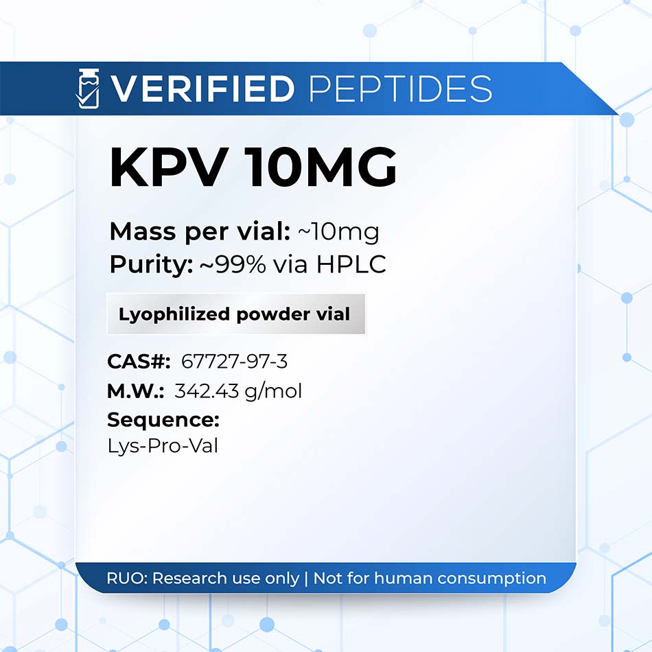
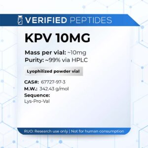
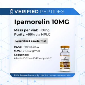
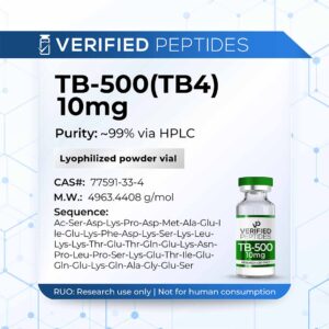
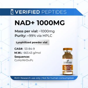
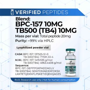
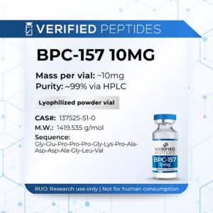
This product is in powder form and is not reconstituted.
All products and materials sold on this site are not for human consumption and subject to our Terms and Conditions.
SHIPS TODAY Order by 1:00 PM EST
FREE SHIPPING For orders over $200
ICE PACK SHIPPING Styrofoam box shipping available
Subject to our Terms and Conditions. This material is sold for laboratory research use only. Not for human consumption, animal, or medical use.
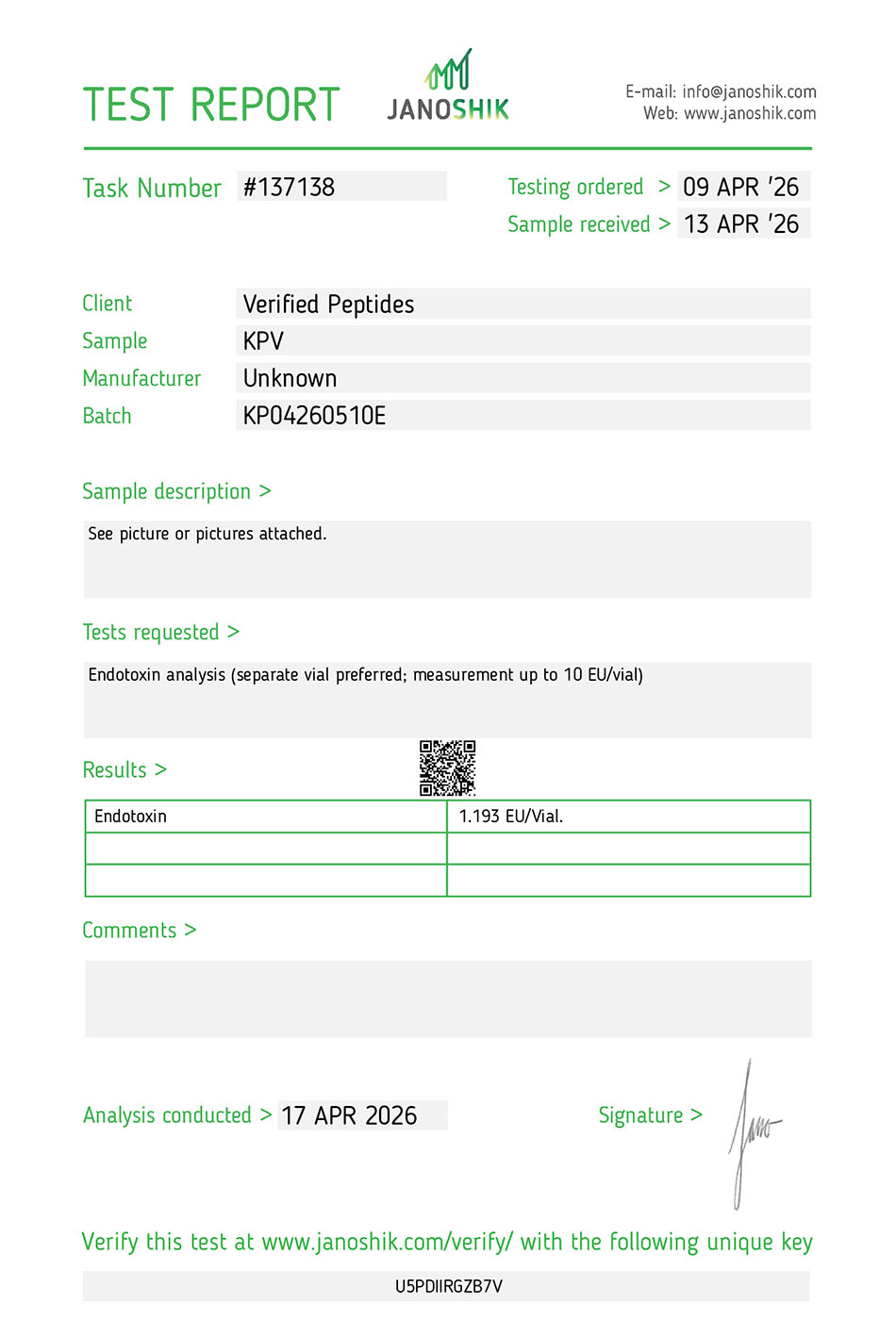
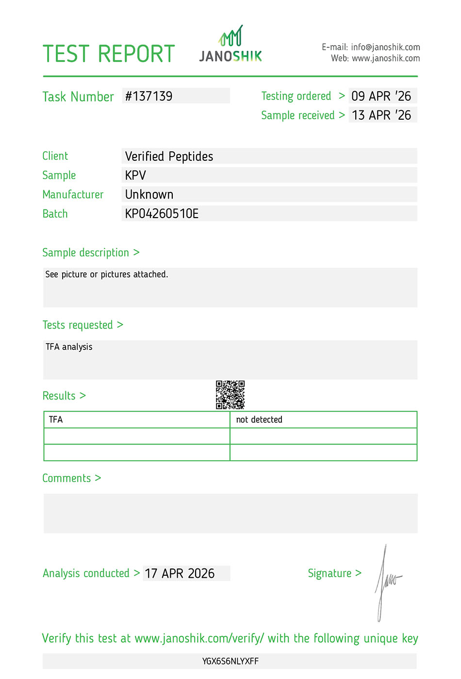
Why isn’t there more information on Verified Peptides KPV peptides?
Due to the legal landscape of peptides and research products, providing information that may imply anything beyond laboratory research use is a legal liability. We’re an expert biotechnology company that provides high quality peptides and products for purchase to advance scientific research in this field.
Why Choose Verified Peptides?
The industry's most trusted source for research peptides
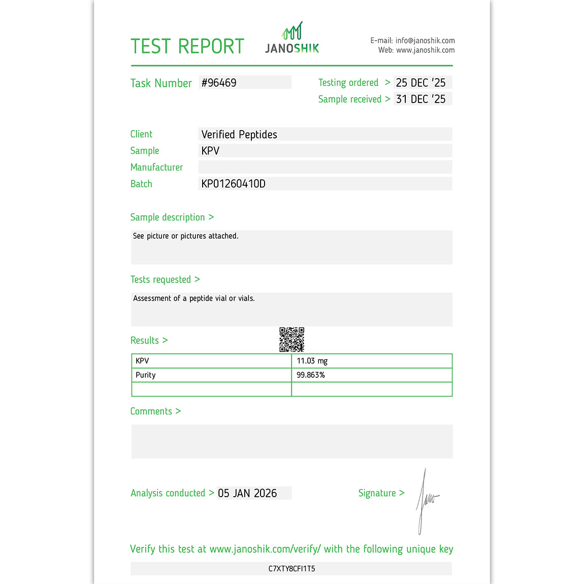
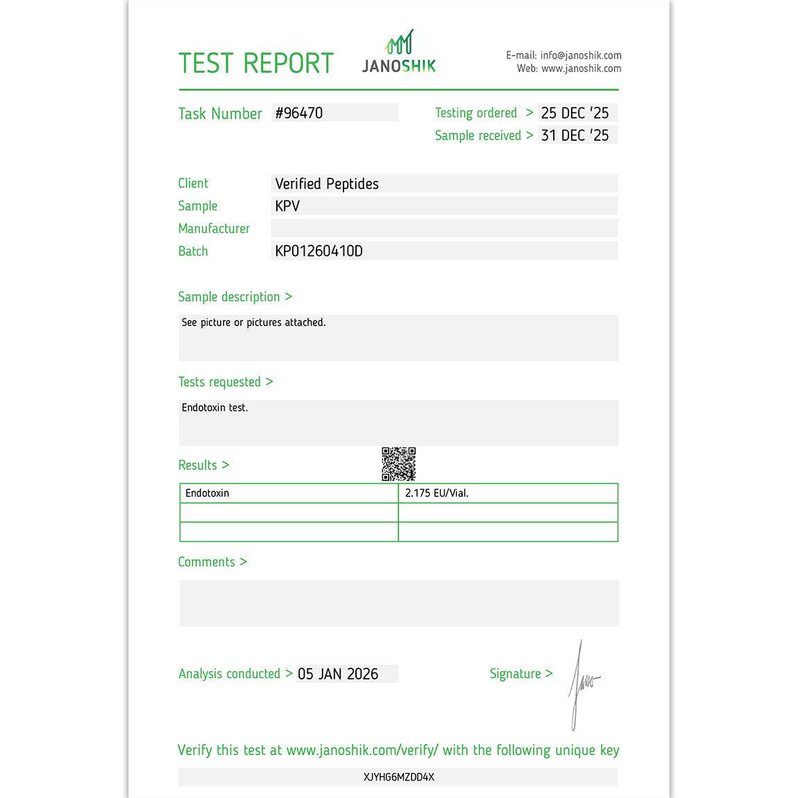
The #1 Leader In Peptide Testing 400+ Certificates of Analysis
What does this mean for you? You can be sure we don't cut corners on testing every single batch. Visit our Lab Reports page.
A Long Standing Trusted Company 6+ Years of Peptides
We pioneered third party lab testing and have stood the test of time. Our first lab report dates back to 2019 proving our track record.
The Best $/mg Prices At The Highest Quality
You won't find this degree of testing and quality at this price.
Peptides Tested For More Than Just Purity
We conduct an array of tests via HPLC and other methods for our peptides: purity, weight, endotoxins(LPS), sterility (bacteria & mold/yeast), and TFA content.
This is a first class operation. Quality products and the best customer service. There may be many similar type of companies out there, but this one is trusted and affordable. No price gouging! I highly recommend this company!
Verified Peptides is hands down my favorite website for these products. I can always count on the products to be legitimate and effective, all while being affordable. Will continue to give them my business.
KPV is a tripeptide (lysine-proline-valine) corresponding to positions 11-13 of α-melanocyte-stimulating hormone (α-MSH), a member of the melanocortin peptide family derived from the pro-opiomelanocortin (POMC) precursor protein. Despite being only three amino acids long—representing the COOH-terminal fragment of the full 13-amino acid α-MSH molecule—KPV retains the potent anti-inflammatory and antipyretic signaling activity associated with the complete peptide. This remarkable functional preservation in such a compact structure has made KPV a valuable research tool for investigating melanocortin biology, particularly in the context of immune regulation. Prior research established that KPV can modulate inflammatory responses in multiple systems, making it especially relevant for studying how the brain's resident immune cells—microglia—regulate their inflammatory output. The peptide's small size, stability, and preserved bioactivity position it as a tractable model for understanding the endogenous mechanisms that balance immune activation and inflammatory control in both peripheral and central nervous system contexts.
KPV Peptide Research Studies
Peer-reviewed scientific research findings
📚 Peer-Reviewed Study
Small Peptide, Big Impact: How KPV Suppresses Proinflammatory Cytokines and Reveals an Autocrine Brake on Microglial Activation
▼
Overview of the Study
This study, published by Delgado and colleagues in the Journal of Leukocyte Biology (1998), investigated whether melanocortin peptides—specifically α-MSH (1-13), KPV (α-MSH 11-13), and ACTH (1-24)—could modulate the inflammatory response of activated murine microglia, the resident macrophages of the central nervous system. Microglia are critical CNS immune cells that, when activated, produce proinflammatory mediators including tumor necrosis factor-α (TNF-α), interleukin-6 (IL-6), and nitric oxide (NO). Excessive production of these cytokines has been implicated in tissue damage associated with multiple sclerosis, Alzheimer's disease, HIV-associated neurological disorders, and other CNS pathologies.
The research addressed three interconnected questions: First, can these melanocortin peptides inhibit TNF-α, IL-6, and NO production by activated microglia, and does elevated intracellular cyclic adenosine monophosphate (cAMP) mediate this effect? Second, do microglia themselves produce and secrete α-MSH, which would enable an autocrine anti-inflammatory feedback circuit? Third, does neutralizing endogenous α-MSH worsen the inflammatory response, providing functional evidence that the peptide acts as a natural brake on microglial activation? The findings demonstrated robust suppression of all three inflammatory mediators by melanocortin peptides, confirmed microglial production of α-MSH, and revealed that neutralizing this endogenous peptide significantly amplified inflammatory output—establishing an autocrine regulatory mechanism in brain-resident immune cells. All findings summarized here derive from in vitro cell culture experiments using immortalized murine microglial cells.
Why KPV and Melanocortin Signaling in Microglia Matter
Microglia represent the principal source of proinflammatory cytokines within the CNS. Under resting conditions, these cells maintain a quiescent, surveillance phenotype with downregulated antigen expression. However, upon activation by pathogens, tissue damage, or protein aggregates, microglia transform into potent producers of inflammatory mediators. While this response is protective in acute contexts, excessive or prolonged microglial activation drives neuroinflammation that damages neurons and contributes to disease progression in multiple neurological conditions.
The melanocortin peptide family, particularly α-MSH, had previously demonstrated anti-inflammatory properties in peripheral immune cells. Research had established that α-MSH could suppress cytokine production by peripheral monocytes and macrophages, partly through an autocrine feedback mechanism where activated immune cells produce α-MSH to self-limit their inflammatory response. Whether this regulatory system extended to CNS-resident immune cells remained unknown. KPV, as the COOH-terminal tripeptide of α-MSH, had shown retention of anti-inflammatory activity in peripheral systems despite its dramatically reduced size, making it a valuable tool for mechanistic investigation. Understanding whether melanocortin signaling could modulate microglial inflammatory output—and whether microglia possessed the autocrine α-MSH circuit observed in peripheral macrophages—would provide important insights into endogenous mechanisms for controlling neuroinflammation. This knowledge could inform research strategies for managing CNS inflammatory conditions where microglial overactivation contributes to pathology.
Experimental Design and Methodology
The study employed the N9 murine microglial cell line, an immortalized cell line derived from embryonic mouse brain cultures that retains key functional properties of primary microglia. Cells were plated at 2×10⁵ cells/mL and activated using a well-established inflammatory stimulus: lipopolysaccharide (LPS, 10 ng/mL) plus interferon-γ (IFN-γ, 1 U/mL) for 24 hours. This combination robustly induces cytokine and nitric oxide production in macrophage-lineage cells, creating a strong inflammatory phenotype suitable for testing anti-inflammatory interventions.
Three melanocortin peptides were tested: α-MSH (1-13, the full-length melanocyte-stimulating hormone), KPV (positions 11-13 of α-MSH), and ACTH (1-24, a truncated form of adrenocorticotropic hormone). Peptides were tested primarily at micromolar concentrations (1, 10, 25, 50, and 100 µM), added to culture wells 10 minutes before LPS plus IFN-γ challenge. Cell viability assessed by trypan blue exclusion consistently exceeded 98% across all conditions, confirming that observed effects reflected pharmacological modulation rather than cellular toxicity. Primary outcome measures included TNF-α production (measured by bioassay using L929 cells), IL-6 levels (quantified by ELISA), and nitric oxide production (assessed via Griess nitrite assay as a proxy for NO synthesis). Mechanistic investigations measured intracellular cAMP accumulation and employed Northern blot analysis to examine TNF-α and inducible nitric oxide synthase (iNOS) mRNA levels. To test the autocrine hypothesis, α-MSH concentrations in conditioned supernatants were measured by radioimmunoassay, and a neutralization experiment used rabbit anti-α-MSH antibody (1:250 dilution) to block endogenous peptide activity before and during inflammatory stimulation. These parameters describe in vitro experimental conditions in cultured mouse microglial cells and do not translate to dosing or applications in other contexts.
Key Findings — Robust Suppression of TNF-α, IL-6, and NO
At 10 µM—the concentration producing peak inhibitory activity—α-MSH (1-13) reduced LPS plus IFN-γ-stimulated TNF-α, IL-6, and nitrite production by approximately 43%, 31%, and 42%, respectively. The dose-response relationship followed a U-shaped (non-monotonic) pattern consistent with earlier reports on α-MSH biology, where both very low and very high concentrations showed reduced efficacy compared to intermediate concentrations.
KPV (11-13) demonstrated comparable or slightly superior efficacy, achieving reductions of approximately 45%, 50%, and 40% for TNF-α, IL-6, and NO, respectively. This finding was particularly significant as it demonstrated that the three-amino acid COOH-terminal fragment retained full anti-inflammatory potency despite representing only 23% of the full α-MSH sequence. ACTH (1-24) also produced substantial inhibition, reducing these three mediators by approximately 38%, 65%, and 41%, respectively, confirming that anti-inflammatory activity extends across multiple members of the melanocortin peptide family. The observation that KPV matched or exceeded the efficacy of the full-length α-MSH molecule, despite its drastically simplified structure, underscored its value as both a research tool and a potentially therapeutically relevant fragment worthy of further investigation.
Suppression Occurs at the Transcriptional Level
Northern blot analysis revealed that the reductions in secreted TNF-α and NO were not simply due to post-translational interference or enhanced degradation of already-produced mediators, but rather reflected suppression at the gene expression level. Melanocortin peptide treatment decreased both TNF-α mRNA and iNOS mRNA in activated microglia, indicating that these peptides were intervening upstream in the inflammatory cascade by preventing transcription of the genes encoding these inflammatory mediators.
This mechanistic insight is important because transcriptional suppression represents a more fundamental level of inflammatory control than simply blocking the activity of already-synthesized proteins. By preventing mRNA production, melanocortin peptides reduce the cellular commitment to inflammatory mediator synthesis, potentially enabling a more complete resolution of the activated state. The parallel suppression of both TNF-α and iNOS transcripts suggests that melanocortin signaling converges on common transcriptional regulatory mechanisms—likely involving cAMP-responsive transcription factors—that coordinately control multiple inflammatory gene programs. This coordinated suppression of inflammatory transcription distinguishes melanocortin peptides from interventions that selectively block individual cytokines or enzymes, positioning them as modulators of the broader inflammatory transcriptional program.
cAMP Signaling: The Central Mechanistic Pathway
Both α-MSH (1-13) and KPV (11-13) produced marked increases in intracellular cAMP accumulation in N9 microglia, and this effect persisted when cells were co-incubated with LPS plus IFN-γ. Critically, the magnitude of cAMP elevation induced by melanocortin peptides was comparable to that produced by forskolin—a direct pharmacological activator of adenylyl cyclase used as a positive control in these experiments. This finding established that cAMP elevation represents a primary downstream consequence of melanocortin peptide engagement in microglial cells.
The mechanistic importance of cAMP elevation is reinforced by convergent evidence from other experimental systems. Multiple independent studies had demonstrated that cAMP-enhancing agents, including the β-adrenergic agonist isoproterenol and the adenylyl cyclase activator forskolin, suppress iNOS expression and TNF-α production in microglial cultures. This body of evidence supports the interpretation that elevated intracellular cAMP acts as a central signaling node linking melanocortin receptor activation to suppression of inflammatory gene transcription. The cAMP second messenger system activates protein kinase A (PKA), which in turn phosphorylates transcription factors including cAMP response element-binding protein (CREB) and potentially interferes with NF-κB activation—transcriptional regulators that control inflammatory gene expression. The finding that KPV elevates cAMP to the same degree as full-length α-MSH, despite potentially engaging different receptor subtypes, suggests that cAMP elevation represents a common downstream pathway through which diverse melanocortin peptides exert anti-inflammatory effects.
Microglia Produce α-MSH: An Autocrine Anti-Inflammatory Circuit
A critical discovery was that microglia themselves produce and release α-MSH when stimulated with inflammatory signals. Cells exposed to LPS, IFN-γ, or the combination released measurable quantities of α-MSH into conditioned culture medium, as detected by radioimmunoassay. This finding established that microglia possess the enzymatic machinery to process POMC precursor protein and generate bioactive α-MSH, extending observations previously made in peripheral monocytes and macrophages into CNS-resident immune cells.
The functional significance of this endogenous α-MSH production was demonstrated through immunoneutralization experiments. When endogenous α-MSH was blocked using anti-α-MSH antibody prior to and during LPS plus IFN-γ stimulation, production of TNF-α, IL-6, and NO increased significantly—by approximately 28%, 26%, and 27%, respectively—compared to cells treated with control IgG at the same dilution. α-MSH levels in supernatants from antibody-treated cells were undetectable, confirming successful neutralization of the endogenous peptide. These results provide compelling functional evidence that activated microglia depend on self-produced α-MSH as an autocrine brake on their own inflammatory response. The inflammatory stimulus that triggers cytokine production simultaneously induces production of the peptide that attenuates it, creating a self-limiting negative feedback loop. This autocrine regulatory circuit represents an intrinsic mechanism for preventing excessive inflammatory responses that could damage surrounding neural tissue. The magnitude of the effect—approximately 25-30% increase in inflammatory mediators when the brake is removed—indicates that this endogenous regulatory system makes a functionally significant contribution to controlling the intensity of microglial inflammatory responses.
Receptor Pharmacology: An Unresolved Question
At the time of this study's publication, five G-protein-coupled melanocortin receptors (MC1R through MC5R) had been identified and cloned. These receptors are distributed in peripheral tissues and throughout the brain, and all couple to adenylyl cyclase to induce intracellular cAMP elevation—consistent with the cAMP increases observed with melanocortin peptide treatment. The receptors show differential expression patterns and ligand preferences: peripheral melanocortin receptors preferentially bind α-MSH, while MC2R (expressed in the adrenal cortex) recognizes only ACTH. Brain melanocortin receptors demonstrate similar affinities for both ACTH and α-MSH.
However, a notable gap in understanding concerned KPV's receptor pharmacology. None of the characterized melanocortin receptors had been shown to directly recognize or bind KPV (α-MSH 11-13). Prior binding studies demonstrated that KPV did not compete with radiolabeled α-MSH for binding at MC1R on melanoma cells, indicating that KPV's receptor interaction profile differs from that of the full-length peptide. This raised an important mechanistic question: if KPV does not bind classical melanocortin receptors in the same manner as α-MSH, how does it produce parallel anti-inflammatory effects and cAMP elevation?
The authors explicitly acknowledged this unresolved issue, noting that the parallel anti-inflammatory effects of α-MSH and KPV across multiple experimental systems might reflect either shared receptor engagement through non-classical binding modes or activation of distinct, as-yet-uncharacterized receptors. Regardless of the receptor question, both peptides elevated cAMP to comparable degrees and produced overlapping suppression of cytokine and iNOS outputs, indicating that whatever molecular mechanism mediates KPV's cellular entry or receptor engagement, its downstream signaling converges on the cAMP pathway that drives anti-inflammatory effects. Resolving KPV's receptor pharmacology remains an important objective for fully understanding the molecular basis of melanocortin anti-inflammatory signaling, though the functional consequences—cAMP elevation and inflammatory suppression—are well-established even without complete receptor identification.
Implications for Understanding Neuroinflammation Control
These findings position the melanocortin peptide system, particularly the KPV fragment, as a potentially important endogenous mechanism for modulating neuroinflammation. Microglia and brain macrophages are the dominant CNS sources of TNF-α, IL-6, and NO—mediators that, while protective in acute responses to infection or injury, become neurotoxic when produced chronically or excessively. The ability of melanocortin peptides to suppress these outputs at the transcriptional level, coupled with the discovery that microglia employ an autocrine α-MSH circuit to self-limit their inflammatory activation, suggests that this system represents a natural regulatory mechanism that the CNS uses to prevent excessive immune activation.
The research findings have particular relevance for neurological conditions characterized by chronic microglial activation and neuroinflammation. In multiple sclerosis, activated microglia contribute to demyelination and axonal damage through excessive cytokine and reactive nitrogen species production. In Alzheimer's disease, microglia activated by amyloid plaques produce inflammatory mediators that exacerbate neuronal dysfunction and death. In HIV-associated neurocognitive disorders, chronically activated microglia driven by viral proteins generate neurotoxic factors contributing to cognitive decline. The demonstration that melanocortin peptides—particularly the remarkably compact KPV tripeptide—can attenuate the key inflammatory outputs implicated in these pathologies suggests potential research applications for understanding how to support or enhance endogenous anti-inflammatory mechanisms.
The autocrine circuit discovery adds another layer of translational relevance. If activated microglia naturally produce α-MSH to self-limit their inflammatory response, then therapeutic strategies might focus on enhancing this endogenous circuit—either by preventing α-MSH degradation, augmenting its production, or supplementing with exogenous peptide—rather than introducing entirely foreign regulatory mechanisms. The finding that immunoneutralization of endogenous α-MSH amplified inflammatory output by approximately 25-30% indicates that this autocrine system makes a functionally meaningful contribution even under basal conditions, suggesting that interventions supporting this pathway could have measurable anti-inflammatory effects.
Discussion and Research Context
This study provided the first comprehensive characterization of melanocortin peptide effects on CNS-resident immune cells, extending observations from peripheral macrophage systems into microglia. The parallel demonstration of direct anti-inflammatory effects (exogenous peptide suppressing cytokines) and autocrine regulatory mechanisms (endogenous peptide production and functional importance) established that melanocortin signaling represents both a potential therapeutic target and an intrinsic regulatory system worth supporting or preserving.
Several aspects of the findings merit emphasis. First, the non-monotonic dose-response relationship—where intermediate concentrations produced maximal effects while very high concentrations showed reduced efficacy—is characteristic of melanocortin peptide biology and may reflect receptor desensitization or engagement of inhibitory pathways at high concentrations. This pattern underscores the importance of dose optimization in any research applications and suggests that more is not necessarily better with melanocortin-based interventions. Second, the preservation of full anti-inflammatory activity in KPV despite its drastic size reduction from the 13-amino acid α-MSH represents both a scientific puzzle (what structural features of these three amino acids are essential?) and a practical advantage (smaller peptides often have better stability and tissue penetration). Third, the transcriptional level of suppression—decreasing inflammatory gene mRNA rather than simply blocking protein activity—suggests that melanocortin signaling may enable more complete resolution of inflammatory activation compared to interventions targeting downstream effectors.
Important limitations must be acknowledged. All data derive from a single immortalized cell line (N9 murine microglia) cultured under artificial in vitro conditions. While this cell line retains key microglial properties, primary microglia freshly isolated from brain tissue might respond differently. The concentrations employed (primarily micromolar) are experimental culture conditions that do not translate to dosing recommendations for any in vivo application. Species differences between mouse and human microglia may exist, potentially limiting direct translation. Most critically, in vitro cell culture cannot replicate the complex multicellular environment of the brain, where astrocytes, neurons, and other cell types participate in regulating microglial activation. The receptor pharmacology questions surrounding KPV remain unresolved, limiting mechanistic understanding even as functional effects are well-documented.
Conclusion
In this controlled in vitro study, melanocortin peptides α-MSH (1-13), KPV (α-MSH 11-13), and ACTH (1-24) significantly suppressed production of the proinflammatory mediators TNF-α, IL-6, and nitric oxide by LPS plus IFN-γ-activated murine microglia. The suppression occurred at the transcriptional level, with decreased TNF-α and iNOS mRNA, and was associated with marked elevation of intracellular cAMP—suggesting that cAMP-dependent signaling pathways mediate the anti-inflammatory effects. The compact tripeptide KPV demonstrated efficacy comparable to or exceeding the full-length α-MSH molecule, indicating that the COOH-terminal sequence retains complete anti-inflammatory potency.
Critically, the study revealed that activated microglia themselves produce α-MSH, and neutralizing this endogenous peptide significantly amplified inflammatory mediator production—approximately 25-30% increases in TNF-α, IL-6, and NO. This finding established that CNS-resident immune cells possess an autocrine melanocortin regulatory circuit that functions as a natural brake on inflammatory activation, extending observations from peripheral macrophages into brain-resident phagocytes. However, it is crucial to emphasize that all findings reported here arise entirely from in vitro experiments using immortalized mouse microglial cells cultured under defined laboratory conditions. The concentrations tested represent experimental culture parameters and do not translate to dosing guidance for any other context. The effects, mechanisms, receptor pharmacology, and therapeutic relevance of KPV and related melanocortin peptides in human neurological conditions remain undefined without extensive additional research including in vivo studies and eventual clinical investigation. These peptides remain research tools for investigating endogenous anti-inflammatory mechanisms and microglial biology. Nevertheless, the demonstration of robust anti-inflammatory effects, transcriptional-level suppression, cAMP-mediated signaling, and functional autocrine regulation establishes melanocortin peptides—particularly the remarkably potent KPV tripeptide—as compelling subjects for continued investigation in neuroinflammation research.
Frequently Asked Questions
What are melanocortin peptides and how does KPV fit in?
Melanocortin peptides are amino acid sequences derived from the pro-opiomelanocortin (POMC) precursor protein, including α-melanocyte-stimulating hormone (α-MSH) and adrenocorticotropic hormone (ACTH). KPV (lysine-proline-valine) is the COOH-terminal tripeptide corresponding to positions 11-13 of the 13-amino acid α-MSH molecule. Despite representing only 23% of the full-length peptide, KPV retains complete anti-inflammatory and antipyretic activity, making it a compact and valuable research tool for studying melanocortin signaling.
Which proinflammatory cytokines did KPV reduce in microglia models?
In LPS plus IFN-γ-activated N9 murine microglia, KPV (α-MSH 11-13) at 10 µM reduced TNF-α by approximately 45%, IL-6 by approximately 50%, and nitric oxide (measured as nitrite, reflecting iNOS activity) by approximately 40%. Northern blot analysis confirmed corresponding decreases in TNF-α mRNA and iNOS mRNA, demonstrating that suppression occurred at the gene transcription level rather than simply blocking already-produced proteins.
How does cAMP signaling relate to KPV's anti-inflammatory effects?
Both α-MSH (1-13) and KPV (11-13) induced substantial intracellular cAMP accumulation in microglia, reaching levels comparable to those produced by forskolin—a direct adenylyl cyclase activator. Because multiple independent studies had shown that cAMP-raising agents suppress iNOS expression and TNF-α production in microglia, the investigators concluded that cAMP elevation is the central signaling mechanism linking melanocortin receptor engagement to reduced inflammatory mediator output, likely through activation of protein kinase A and modulation of inflammatory transcription factors.
Do microglia produce α-MSH, and why does that matter?
Yes. Microglia stimulated with LPS and/or IFN-γ released measurable α-MSH into culture supernatants as detected by radioimmunoassay. When this endogenous peptide was immunoneutralized using anti-α-MSH antibody, TNF-α, IL-6, and NO production increased by approximately 26-28% compared to control conditions. This demonstrates that microglia rely on self-produced α-MSH as an autocrine brake on their own inflammatory activation—a negative feedback loop where the inflammatory stimulus simultaneously triggers both cytokine production and the peptide that attenuates it, creating an intrinsic mechanism for preventing excessive inflammation.
Are KPV's receptor targets the same as classical melanocortin receptors?
This remains an unresolved question. Five G-protein-coupled melanocortin receptors (MC1R-MC5R) have been characterized and all couple to adenylyl cyclase to elevate cAMP. However, there was no evidence at the time of this study's publication that known MC receptors directly recognize or bind KPV. Prior research showed KPV did not compete with radiolabeled α-MSH for MC1R binding, suggesting its receptor pharmacology differs from full-length α-MSH. The parallel anti-inflammatory effects may reflect either non-classical binding to known receptors or engagement of distinct, uncharacterized receptors—a question requiring further investigation.
What do these findings suggest about balancing brain immunity?
The findings suggest the CNS possesses endogenous melanocortin-based mechanisms for moderating microglial inflammatory activity. Both exogenously applied KPV and endogenously produced α-MSH suppress TNF-α, IL-6, and NO output through cAMP-dependent pathways. The discovery of a functional autocrine circuit—where microglia produce α-MSH that self-limits their inflammatory response—reveals an intrinsic regulatory system that may be relevant for preventing excessive neuroinflammation. This positions the melanocortin system as a research target for understanding how the brain naturally controls immune activation.
How do melanocortin peptides alter gene expression in activated microglia?
Northern blot analysis revealed that melanocortin peptides reduced TNF-α mRNA and iNOS mRNA levels in activated microglia, indicating transcriptional-level suppression rather than post-translational interference. The cAMP elevation induced by these peptides likely activates protein kinase A, which phosphorylates transcription factors including CREB and potentially interferes with NF-κB activation—key regulators that control inflammatory gene expression. This coordinated suppression of multiple inflammatory transcripts distinguishes melanocortin peptides from interventions targeting individual cytokines or enzymes.
Why is the autocrine α-MSH circuit important for neuroinflammation?
The autocrine circuit creates a self-limiting negative feedback loop where the same inflammatory stimuli that drive cytokine production also trigger α-MSH release that attenuates the response. When this endogenous brake is removed through antibody neutralization, inflammatory outputs increase by approximately 25-30%—demonstrating that the autocrine system makes a functionally significant contribution to controlling microglial activation intensity. This suggests that the integrity and function of this melanocortin regulatory circuit may influence the magnitude and duration of neuroinflammatory responses in CNS pathology.
What concentrations of KPV were tested in this study?
KPV was tested at 1, 10, 25, 50, and 100 µM concentrations in N9 microglial cell cultures, with 10 µM producing peak inhibitory effects on TNF-α, IL-6, and NO production. The dose-response curve showed a U-shaped (non-monotonic) pattern where intermediate concentrations were most effective. These represent in vitro experimental culture conditions in immortalized mouse cells and do not translate to dosing recommendations for any other context or application.
Is this in vitro research the same as effects in living organisms or humans?
No. All findings come from controlled in vitro experiments using the N9 immortalized murine microglial cell line cultured under defined laboratory conditions. While this system provides valuable mechanistic insights, it cannot replicate the complex multicellular environment of the brain where astrocytes, neurons, blood-brain barrier, and other factors influence microglial behavior. Species differences between mouse and human microglia may exist. The effects, optimal conditions, safety, and relevance of KPV in living organisms or human neurological conditions remain undefined without extensive additional research. These peptides remain research tools requiring substantial further investigation.
📚
Study Reference
Delgado R, Carlin A, Airaghi L, et al. Melanocortin peptides inhibit production of proinflammatory cytokines and nitric oxide by activated microglia. J Leukoc Biol. 1998;63(6):740-745. doi:10.1002/jlb.63.6.740