
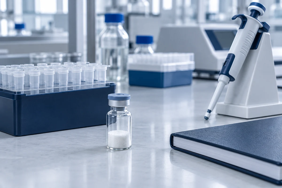
Short answer
To reconstitute a lyophilized peptide, let the sealed vial reach room temperature, collect the powder at the bottom, and add a measured volume of a solvent suited to the sequence slowly down the vial wall. Mix gently until the solution is clear, then divide the stock into single-use aliquots, label them, and store them frozen at −20 °C or colder unless the supplier specifies otherwise.
Below: supplies, planning, the step-by-step method, solvent choice and its effect on stability, concentration math, proteins and reference standards, storage, aseptic handling, and troubleshooting.
What Is Peptide Reconstitution and Why Does It Matter?
Peptide reconstitution is the process of dissolving a lyophilized (freeze-dried) peptide in a measured volume of solvent to make a stock solution of known concentration. Peptides are supplied dry because they are generally far more stable as a powder; once in solution, their shelf life is limited.1, 2
The step looks simple, but it is where many downstream problems start. A peptide that is only partly dissolved, a concentration that ignores net peptide content, a solvent that slowly degrades the sequence, or a stock contaminated during handling can all show up later as noise, drift, or an apparent experimental effect.
It helps to think of reconstitution as choosing the environment the peptide will sit in until it is used, not as a one-time event. The solvent, pH, concentration, and storage conditions together decide how long the stock stays reliable. For background on the dry form, see what a lyophilized peptide is.
Supplies for Reconstituting Peptides in the Lab
Gather everything before the vial is opened, so it stays open for as short a time as possible.
- The peptide vial with its Certificate of Analysis (COA) and any product datasheet, which give the peptide mass, net peptide content, and sometimes a recommended solvent or concentration.3
- A first-choice solvent, usually sterile water or a sterile, assay-compatible buffer, plus dilute acetic acid, dilute ammonium bicarbonate or ammonium hydroxide, and DMSO on hand for sequences that need them.2, 4
- Calibrated micropipettes sized to the volumes you plan to measure, with sterile, low-retention or filtered tips.
- Sterile, low-binding tubes or vials for aliquots, and freezer-safe labels.
- An appropriate aseptic workspace, 70% ethanol, and powder-free gloves. A clean bench and a biosafety cabinet are not interchangeable for personnel protection; use the workspace specified by the laboratory’s risk assessment.5
- A desiccator, or a sealed container with desiccant, for warming cold vials before opening.6
- A centrifuge and holder compatible with the vial, if needed to collect powder at the bottom.7
- Optional: a sonicating water bath, pH paper or a pH meter, a sterile 0.2 µm low-protein-binding membrane filter, and argon or nitrogen for oxygen-sensitive sequences.1
Before You Start: Plan the Stock Solution
Plan three things before adding any solvent: what the supplier recommends, what concentration you want, and whether the peptide is likely to dissolve in your first-choice solvent.
Read the datasheet and COA first. If the supplier specifies a solvent or a reconstitution concentration, start there; protocols for lyophilized proteins and standards in particular are written around the supplier’s stated conditions.7, 8 Check whether the COA reports net peptide content or an assayed peptide amount; if that information is missing, confirm it with the supplier.3
Choose a concentration that is easy to work with. A round number such as 1 mg/mL keeps the arithmetic simple, and a stock that is concentrated enough to dilute later, but not so concentrated that working volumes become too small to pipette accurately, is generally the most practical. The calculations section below shows how to check this.
Check the sequence. Net charge and hydrophobicity predict which solvent is likely to work, and cysteine, methionine, or tryptophan residues flag a peptide that needs protection from oxidation.2, 4 If solubility is uncertain, test a small portion of the peptide first rather than committing the whole vial.2, 4
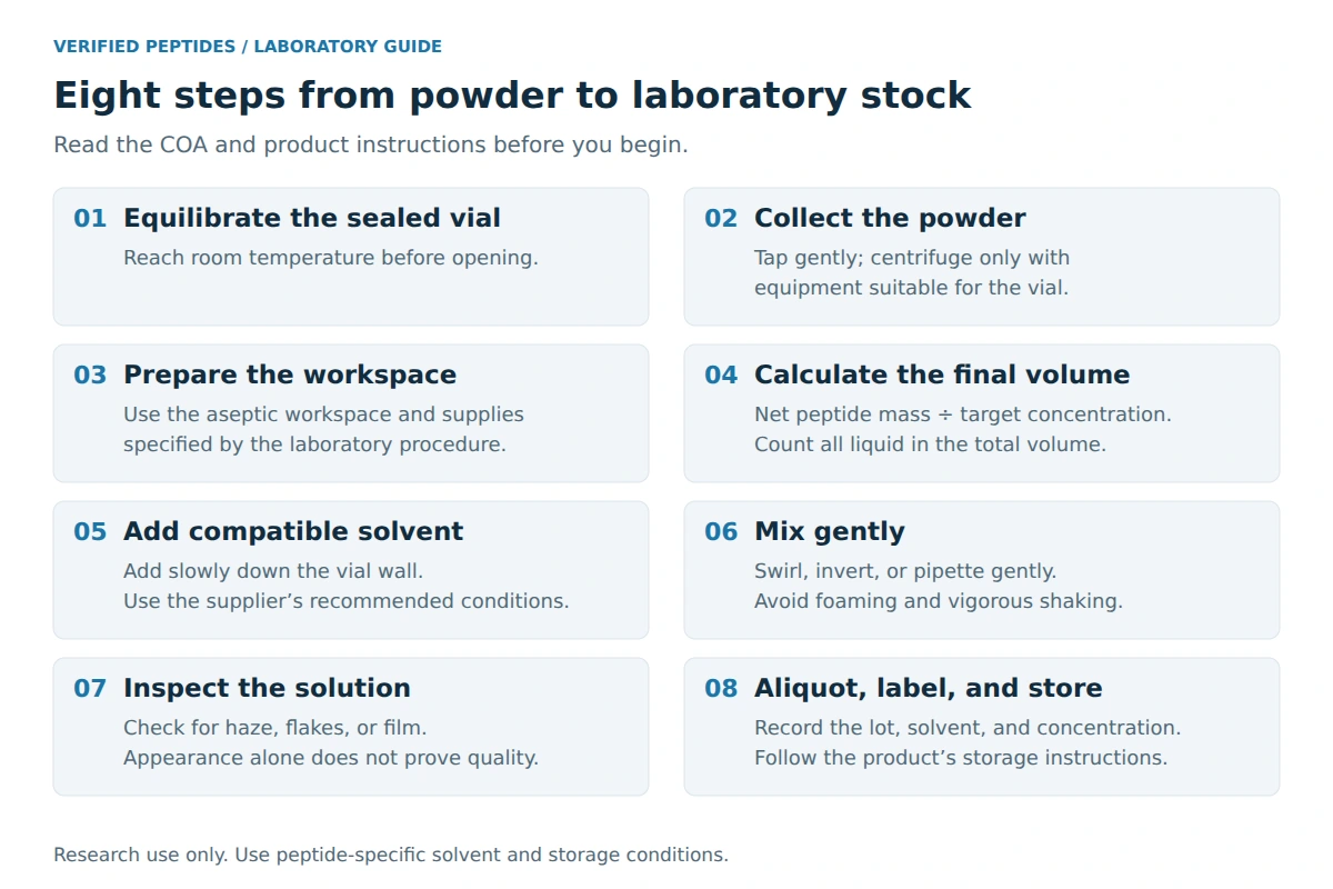
How to Reconstitute Peptides Step by Step
Work through eight steps, keeping the vial open only as long as needed.
- 1Equilibrate the sealed vial Move the vial from cold storage and let it reach room temperature before opening, ideally in a desiccator. Peptides are often hygroscopic, and opening a cold vial lets moisture condense on the powder.1, 4, 6
- 2Collect the powder Gently tap the sealed vial, or briefly centrifuge it with compatible equipment, so the lyophilized material sits at the bottom; powder can cling to the stopper or walls after shipping.7
- 3Prepare a clean workspace Wipe the cabinet surface and your gloves with 70% ethanol, wipe the vial’s septum or cap, and set out sterile tips and tubes.5
- 4Calculate the solvent volume Final solution volume equals net peptide mass divided by the target concentration. For 5 mg of net peptide at 1 mg/mL, prepare a total final volume of 5 mL, including any initial co-solvent. Confirm the container has enough capacity.
- 5Add solvent down the wall Pipette slowly against the inside wall of the vial rather than straight onto the powder. This limits splashing, foaming, and local pockets of very high concentration.
- 6Mix gently Swirl, roll, or invert the vial, or pipette gently up and down. Avoid vigorous shaking, which can cause foaming and denaturation.7 Many peptides dissolve within minutes; for lyophilized proteins, one protocol allows 15 to 30 minutes at room temperature with gentle agitation.7
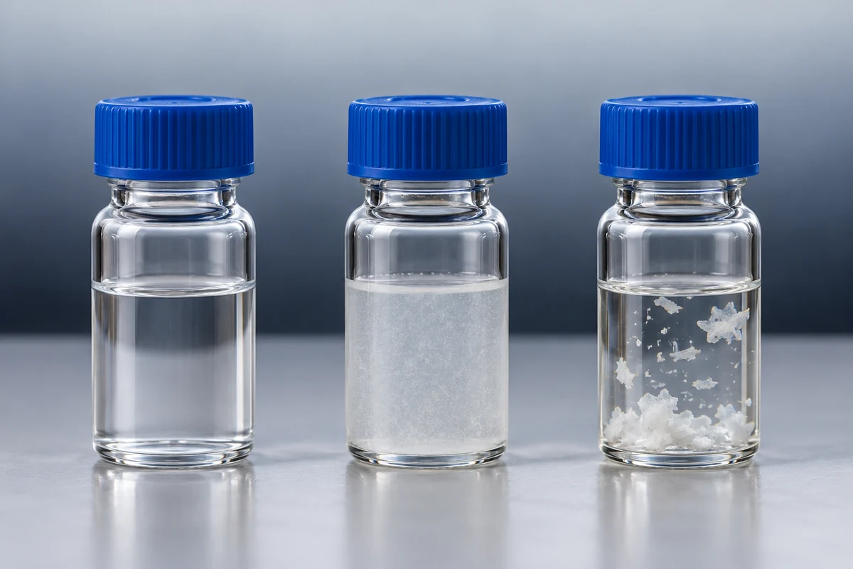
- 7Inspect the solution Check the vial against light and dark backgrounds. A fully reconstituted solution is generally clear and free of flakes or film; if not, see the troubleshooting section.
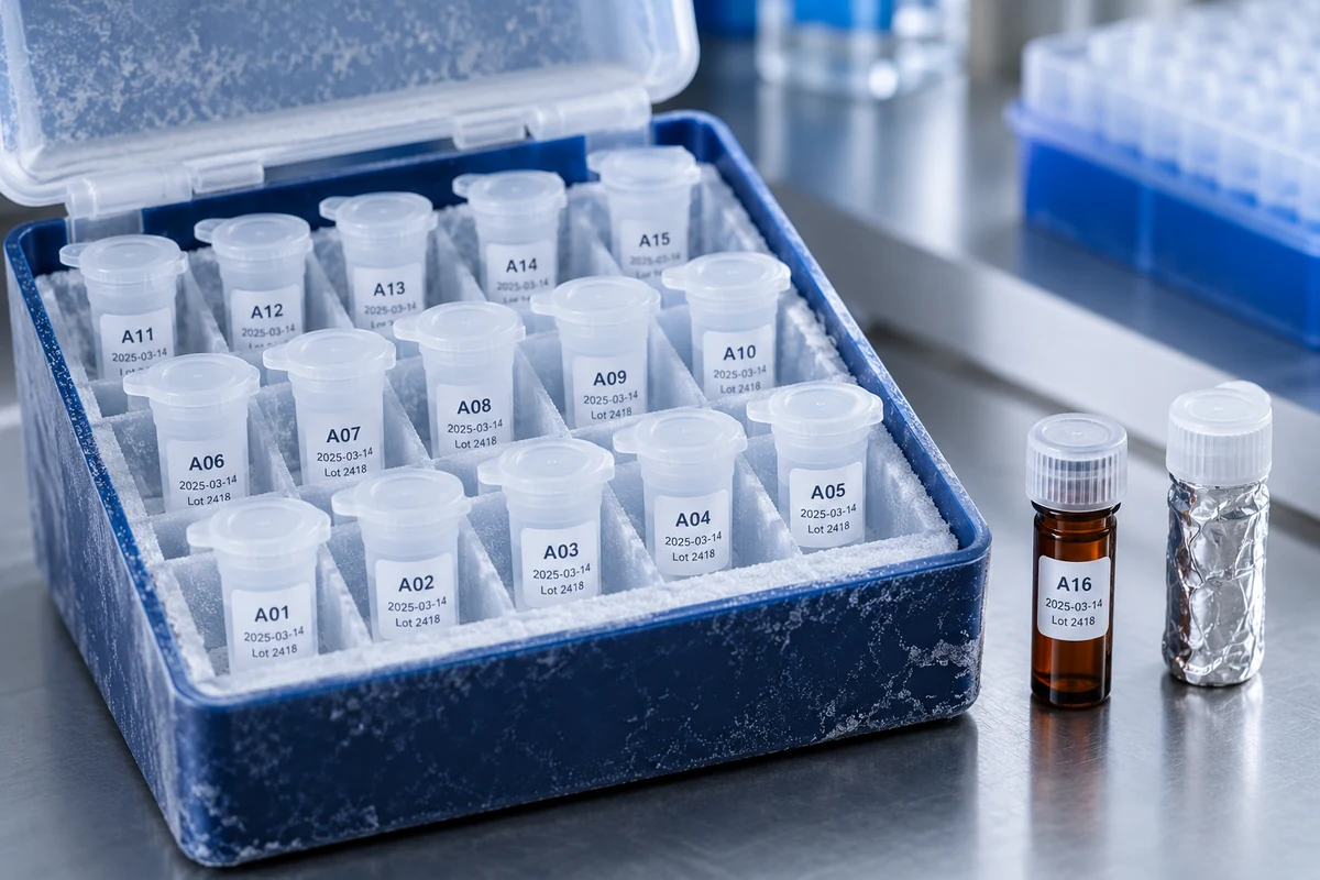
- 8Aliquot, label, and store Divide the stock into single-use tubes, label each with the peptide, lot, solvent, concentration, date, and initials, and store aliquots under the product-specific conditions.1
Can You Vortex a Peptide Solution?
Generally, it is better not to. Gentle swirling or pipetting is the default in supplier protocols, and one protein supplier states plainly not to vortex.8 Some small, robust peptides tolerate brief low-speed vortexing, but unless the datasheet says it is acceptable, gentle mixing is the safer choice.
Choosing the Best Solvent for Peptide Reconstitution
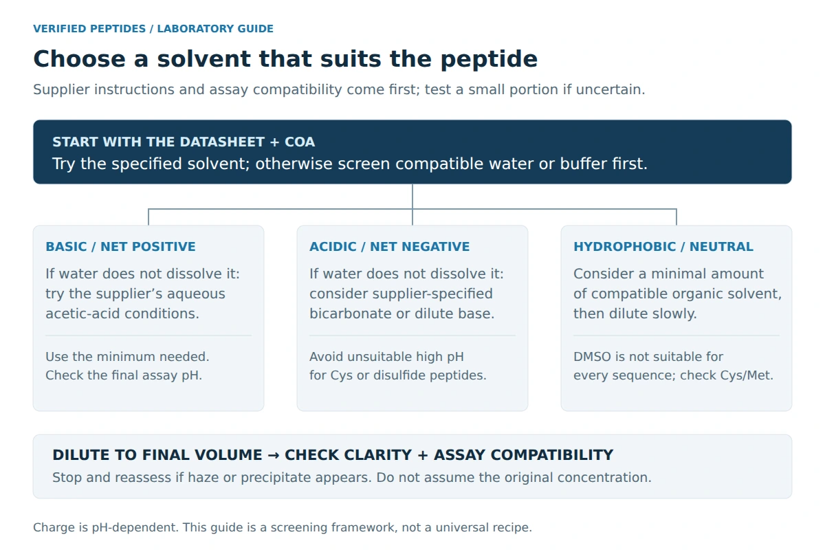
The best solvent is the gentlest one that fully dissolves the peptide and suits the assay. That generally means sterile water or a sterile buffer first, then a dilute acid, a dilute base, or an organic co-solvent, depending on the peptide’s net charge and hydrophobicity.2, 4
For a rough solvent-selection screen, compare basic residues (Lys, Arg, His) with acidic residues (Asp, Glu), and account for free or blocked termini. Actual charge depends on pH, especially for histidine; this count is not an exact net-charge calculation.2 A positive total suggests a basic peptide, a negative total an acidic one, and a total near zero with many hydrophobic residues suggests the peptide will need an organic co-solvent.
| Peptide type | Commonly tried first | If it does not dissolve | Watch for |
|---|---|---|---|
| Basic (net positive) | Sterile water | A small amount of the aqueous acetic-acid solution specified in the supplier’s protocol, then dilute to final volume2, 4 | Keep the final acid level compatible with the assay |
| Acidic (net negative) | Sterile water or PBS | A small amount of 0.1 M ammonium bicarbonate or dilute ammonium hydroxide, then dilute2, 4 | Avoid high pH with free cysteine or disulfide bonds4, 6 |
| Hydrophobic or neutral | A minimal volume of DMSO, DMF, or acetonitrile | Dilute slowly with water or buffer under gentle mixing2, 4 | Cys and Met sequences are unstable in DMSO2 |
| Short or highly charged | Sterile water | Usually dissolves readily | Sequence-specific solubility, pH, and assay compatibility |
Change the solvent in small steps, and add the minimum acid, base, or organic solvent needed before diluting to the final volume. For a deeper look, see choosing solvents for hydrophobic and hydrophilic peptides.
Sterile Water vs. Bacteriostatic Water
For most laboratory work, sterile water or a sterile buffer, combined with single-use aliquots, is the simpler and more controllable choice. Bacteriostatic water contains an antimicrobial preservative, commonly benzyl alcohol, which adds another chemical to the system.
| Property | Sterile water | Bacteriostatic water |
|---|---|---|
| Composition | Water only, no additives | Water with a preservative, commonly benzyl alcohol |
| Assay compatibility | Adds no preservative; pH, ionic strength, and water quality still matter | The preservative may interfere with some cell-based or enzymatic assays |
| Microbial protection | None once opened | Inhibits growth of some bacteria, but does not sterilize a contaminated solution |
| Typical lab use | Stocks that are aliquoted and frozen promptly | Workflows where the preservative is known to be acceptable |
A preservative is not a substitute for aseptic technique, and neither diluent rescues a solution that has become contaminated. Whichever is used, record it on the label.
Saline and Buffered Solutions (PBS)
Buffers make sense when the assay needs a defined pH or ionic strength, or when the datasheet specifies one. Bachem, for example, suggests PBS at pH 7.0 to 7.4 for many charged peptides when a concentration of 1 mg/mL or less is sufficient.6 The trade-offs are that salt can reduce solubility and promote aggregation for some sequences (see the next section), and phosphate can precipitate with certain peptides or trace metal ions. If a peptide clouds in buffer, a common workaround is to dissolve it in water first, then add concentrated buffer to reach the final strength.
Organic Co-Solvents: DMSO, DMF, and Acetonitrile
For hydrophobic or aggregation-prone sequences, dissolve the peptide in the smallest volume of organic solvent that works, then add water or buffer slowly with gentle mixing.2, 4 Adding the aqueous phase too quickly can cause local precipitation; if the solution starts to turn cloudy, stop and reassess.4 Keep the final organic content as low as the assay requires, since DMSO and acetonitrile can affect cells and enzymes, and choose DMF or acetonitrile over DMSO for sequences containing cysteine or methionine.2
How Solvent Choice Affects Peptide Stability
The solvent decides not only whether a peptide dissolves but how quickly it degrades afterward. In formulation research, pH optimization and buffer selection are generally considered the most practical ways to stabilize peptides in solution.9
pH and Buffer Choice
pH controls several common degradation pathways. Asparagine deamidation generally speeds up at neutral and alkaline pH, while bonds next to aspartate can cleave under acidic conditions, so the most stable pH depends on the sequence.9 For general laboratory storage, several peptide suppliers recommend sterile buffers at about pH 5 to 6.1, 2, 4
Free cysteine is a special case: thiols oxidize rapidly to disulfides above pH 7, so Bachem recommends carefully degassed acidic buffers for these sequences.6 If the datasheet notes the peptide was lyophilized from a dilute acid, or recommends a specific buffer, read that as a strong hint about the pH it tolerates.
Ionic Strength and Aggregation
Salt changes how peptide molecules interact. In pure water, highly charged peptides repel one another; added salt shields those charges, which in some cases lets hydrophobic regions associate so the peptide aggregates or precipitates. Ionic strength is a recognized driver of aggregation, and adjusting pH and ionic strength together is a standard stabilization approach.9 In practice, if a peptide dissolves in water but clouds in PBS, the salt is a likely cause; a lower ionic strength, a lower concentration, or a different buffer often helps.
Additives, Preservatives, and Cryoprotectants
Additives can help or get in the way. Polyols and sugars such as glycerol and sucrose have been shown to slow some degradation pathways and are common cryoprotectants for frozen protein stocks,9 but they raise viscosity and can interfere with some assays. Preservatives such as benzyl alcohol limit microbial growth but add another variable. Carrier proteins such as BSA are sometimes added when diluting proteins to working strength, although one supplier advises against adding carrier to the initial reconstitution unless the COA specifies it.8 Add only what the assay can tolerate, and record every additive on the label.
Solvent Volatility and Evaporation
Volatile solvents such as acetonitrile can slowly evaporate from a poorly sealed tube, even in the refrigerator. That changes both the peptide concentration and the solvent ratio, and a hydrophobic peptide may come out of solution as the organic fraction falls. Use tightly sealing tubes and keep organic-containing stocks capped.
Measuring Accurately: Concentration and Dilution Calculations
Concentration equals net peptide mass divided by final solution volume. For example, 5 mg of net peptide in a final volume of 2.0 mL gives a stock of 2.5 mg/mL, which is the same as 2.5 µg/µL.
If you start from gross powder weight, correct for documented net peptide content. If the COA already reports an assayed peptide amount in mg, use that amount directly; do not correct it a second time. HPLC purity is a separate measurement and must not replace the net-content fraction. A lyophilized powder also contains water, salts, and counter-ions such as trifluoroacetate, so the peptide is only part of the weighed mass.3, 10 If the COA reports 80% net peptide content, 5 mg of powder holds about 4 mg of peptide, and the same 2.0 mL gives about 2.0 mg/mL.
| Calculation | Formula | Example |
|---|---|---|
| Stock concentration | Mass ÷ volume | 5 mg ÷ 2.0 mL = 2.5 mg/mL |
| Net-content correction | Mass × net content ÷ volume | 5 mg × 0.80 ÷ 2.0 mL = 2.0 mg/mL |
| Final volume for a target concentration | Net mass ÷ target concentration | 1 mg ÷ 0.2 mg/mL = 5 mL final solution |
| Molar concentration | (mg/mL ÷ molecular weight) × 1,000,000 = µM | 2.0 mg/mL at 1,000 g/mol = 2,000 µM |
| Working dilution | C1 × V1 = C2 × V2 | 2.5 mg/mL × V1 = 0.1 mg/mL × 1 mL, so V1 = 40 µL stock + 960 µL buffer |
Use the same measurement labels from start to finish, and double-check every label (mg, µg, mg/mL, µM) at each step; a slip between them produces a tenfold or thousandfold error. To run these numbers quickly, use the peptide reconstitution calculator, which also shows where a volume falls in your pipette’s range.
Choosing Volumes Your Pipettes Can Measure
Pipettes are generally most accurate near their maximum capacity, and accuracy drops off in the lower part of the range.11 INTEGRA suggests about 35% to 100% of capacity as a preferred working range. This is a selection guideline, not a universal pass/fail limit; check your instrument’s specified range and performance at the actual volume.12 Very small volumes also lose a proportionally larger share to tube walls and tips, which is why protein suppliers advise aliquots of at least 10 to 20 µL.7, 8
If a working volume falls outside your pipette’s suitable operating range, prepare a more dilute stock or an intermediate dilution. For example, 10 µg from a 2.5 mg/mL stock is only 4 µL; diluting 100 µL of stock to 1 mL gives 0.25 mg/mL, and 10 µg is then a comfortable 40 µL.
How to Reconstitute Lyophilized Proteins, Controls, and Standards
Lyophilized proteins follow the same core steps, with more emphasis on gentleness and on the supplier’s instructions: collect the powder by centrifugation, reconstitute in exactly the solvent and concentration given on the datasheet or COA, mix by gentle pipetting or inversion rather than vortexing, and allow 15 to 30 minutes at room temperature with gentle agitation.7, 8
Proteins differ from short peptides in that their activity depends on folded structure, so foaming, shear, and freeze-thaw cycles are generally more damaging. If flakes or particulates remain, one protocol suggests mixing for a couple of hours at room temperature and then overnight at 4 °C before concluding the protein will not dissolve.7
Reconstituting Lyophilized Controls and Reference Standards
For a lyophilized control or standard, follow the datasheet exactly. These products are formulated so that a specified volume of a specified diluent gives a stated concentration, with wording such as “reconstitute in 0.5 mL of deionized water to obtain a 100 µM solution.” Use a calibrated pipette for the full volume, let the vial stand for any time stated, mix gently, and record the lot and reconstitution date.
Changing the volume changes the assigned value, and the result is no longer comparable with the standard curve or with earlier runs. Some reference standards are assigned a biological activity rather than a mass; in that case, keep every label and calculation in the same activity terms the datasheet uses, and apply an activity-to-mass conversion only where the datasheet provides one.
Common Mistakes to Avoid During Reconstitution
Most reconstitution problems come from a handful of avoidable mistakes, and most of those come from rushing.
| Mistake | Why it matters | Better practice |
|---|---|---|
| Opening a cold vial | Condensation adds moisture to a hygroscopic powder | Warm the sealed vial first, ideally in a desiccator |
| Skipping the datasheet | Supplier-specified solvents or concentrations are missed | Read the COA and datasheet before adding anything |
| Dissolving everything in an untested solvent | The whole sample may be lost | Test a small portion first |
| Adding solvent straight onto the powder | Splashing, foaming, and local high concentration | Pipette slowly down the vial wall |
| Shaking or vortexing hard | Foaming, aggregation, and denaturation | Swirl, roll, or pipette gently |
| Using a partly dissolved solution | The real concentration is unknown | Confirm the solution is clear and uniform |
| Ignoring net peptide content | Concentration is overstated | Correct using the COA |
| Mixing up mg, µg, and µM | Tenfold to thousandfold errors | Use one convention and double-check labels |
| Repeatedly thawing one stock | Degradation and contamination | Freeze single-use aliquots |
| Vague or missing labels | Mix-ups and lost traceability | Label peptide, lot, solvent, concentration, and date |
Storage and Stability of Reconstituted Peptides
Prepare single-use aliquots and follow the peptide’s specified storage conditions. Many supplier guides recommend frozen storage at −20 °C or colder for suitable peptide solutions, but freezing is not appropriate for every formulation. Peptides in solution are much less stable than dry powder, and several suppliers advise against storing peptides in solution at all when it can be avoided.1, 6
| Form | Commonly recommended | Notes |
|---|---|---|
| Lyophilized, long term | −20 °C or colder, tightly sealed2, 6 | Bachem notes lower temperatures are preferred |
| Lyophilized, short term | Refrigerator, about 4 °C6 | Warm to room temperature before opening |
| Solution, in use | 2 to 8 °C for short periods | For recombinant proteins, one supplier allows up to about a week8 |
| Solution, longer term | Single-use aliquots frozen at −20 °C or colder1, 6 | Bachem notes frozen peptide solutions may be kept for a few weeks6 |
Stability is sequence-specific, so read these as general guidance and follow any datasheet values. Sequences containing asparagine, glutamine, cysteine, methionine, or tryptophan are generally less stable in solution.2
Each freeze-thaw cycle can degrade a peptide, and repeated opening invites contamination, which is why single-use aliquots are the core of every storage plan.1, 4 Where frozen storage is suitable, thaw an aliquot once and keep it at a temperature that preserves solubility and stability during use. Avoid refreezing unless the laboratory has validated that procedure. See freeze-thaw cycles and peptide degradation.
Protecting Peptides From Light and Oxygen
Sequences containing cysteine, methionine, or tryptophan are prone to oxidation and need extra care.4, 6 Practical steps include using degassed solvents, purging buffers with argon or nitrogen,1 keeping headspace in storage tubes small, and using amber tubes or foil, since light can drive oxidation of aromatic residues.9 Return unused lyophilized peptide to the freezer tightly sealed; see lyophilized peptide storage: temperature, humidity, and light.
Aseptic Handling to Protect Peptide Purity
Aseptic technique keeps microbes, proteases, and particulates out of the stock. Use the aseptic workspace specified for your procedure, wipe surfaces and gloves with 70% ethanol, keep containers capped when not in use, and use each sterile tip once.5
- Wear powder-free gloves and wipe them with 70% ethanol before handling vials.
- Wipe vial septa, caps, and tube racks before they go into the work area.
- Never touch the part of a tip or tube that contacts the solution.
- Protect the inside of caps from contact with the work surface, and recap promptly.
- Keep the solution cool while aliquoting, for example on a chilled rack, unless it needs room temperature to stay dissolved.
For cell-based work, use sterile, endotoxin-tested solvents, since sterility and endotoxin are separate concerns; see endotoxin-free peptide prep and water quality.
Sterile Filtering Without Losing Peptide
A suitable, validated 0.2 µm filtration step can reduce bacterial contamination; it does not reliably remove endotoxin or establish the absence of all contaminants.1 The trade-off is adsorption: some peptide binds to the membrane, especially hydrophobic sequences and dilute solutions. To limit losses, choose a low-protein-binding membrane, follow the membrane manufacturer’s wetting and rinsing instructions, use the smallest filter suited to the volume, and filter only when the work requires it. Filtration backs up good technique rather than replacing it; see minimizing contamination risk from reconstitution to final assay.
Troubleshooting Clumping, Cloudiness, and Incomplete Dissolution
Cloudiness, flakes, or a gel usually mean the peptide has not fully dissolved or has aggregated, so the solution’s real concentration is unknown. Change one condition at a time and note what works.
When the Peptide Will Not Dissolve
- Give it time. Let the vial stand at room temperature with occasional gentle swirling.
- Sonicate briefly. Several minutes in a water bath can break up larger particles; short bursts with cooling in between limit heating.2, 6
- Warm gently. Modest warming can improve solubility, but excessive warming should be avoided.4, 6
- Change the solvent strategy. Match the solvent to the net charge, or switch to a minimal organic co-solvent for hydrophobic sequences.2, 4
- Allow extended mixing for proteins. If flakes persist, mix for a couple of hours at room temperature, then overnight at 4 °C.7
- Re-lyophilize as a last resort. If a volatile solvent such as dilute acetic acid or ammonium bicarbonate was the problem, labs with a freeze dryer can sometimes remove it and start again with a better solvent.
When the Solution Turns Cloudy
| What you see | Likely cause | What to try |
|---|---|---|
| Clear at room temperature, cloudy after chilling | Concentration near the solubility limit at lower temperature | Dilute the stock, or prepare a lower-concentration stock next time |
| Cloudy right after adding buffer | Salt or phosphate effects, or a pH shift | Dissolve in water first, then add buffer; check the pH |
| Cloudy during DMSO dilution | Aqueous phase added too quickly | Restart with slower dilution and gentle mixing |
| Faint haze that does not clear | A small amount of insoluble material | Investigate the cause. If a validated method uses centrifugation or filtration, measure peptide recovery and concentration before using the clarified stock |
| Clear stock turns cloudy days later, sometimes with a film or odor | Possible microbial growth | Discard it; do not try to rescue it |
If a peptide still will not cooperate, contact the supplier with the lot number; technical support may know of sequence-specific quirks. See solubility troubleshooting by sequence type.
Final Tips for Consistent Reconstitution
Consistency comes from doing the same thing the same way every time, and writing it down.
- Put the method in a lab SOP, including solvent, volume, mixing, and storage, so everyone who handles the peptide follows the same procedure.
- Record what worked for each sequence, including any troubleshooting steps, for the next reconstitution.
- Change one variable at a time when something goes wrong.
- Keep the COA with your records so every concentration traces back to batch data.
- When results drift, check the stock (age, freeze-thaw history, appearance) before blaming the experiment.
A clear solution is a good sign, but it is not proof of identity or concentration. The batch COA covers identity and purity at release, and HPLC or mass spectrometry can confirm the peptide in solution when it matters.3 See certificates of analysis: what researchers need to know.
Frequently Asked Questions About Peptide Reconstitution
What are lyophilized peptides, and why do they need to be reconstituted?
They are freeze-dried peptide powders, supplied dry because that form is generally far more stable. Reconstitution dissolves them into a stock solution of known concentration so they can be measured, diluted, and used in laboratory work.
How do you reconstitute lyophilized peptides step by step?
Warm the sealed vial to room temperature, collect the powder, prepare a clean workspace, calculate the final volume, add compatible solvent slowly down the vial wall, mix gently, then aliquot, label, and store as specified.
What is the best solvent for reconstituting lyophilized peptides?
It depends on the sequence. Sterile water or an assay-compatible sterile buffer is the usual starting point, with dilute acetic acid for basic peptides, dilute ammonium bicarbonate for acidic peptides, and a minimal volume of DMSO, DMF, or acetonitrile for hydrophobic ones.
Should you use sterile water or bacteriostatic water?
For most laboratory work, sterile water or a sterile buffer is simpler because it avoids an added preservative; its pH and ionic strength still need to suit the assay. Bacteriostatic water contains a preservative, commonly benzyl alcohol, that may interfere with some assays and does not replace aseptic technique.
How do you reconstitute lyophilized proteins?
Centrifuge the vial briefly, add exactly the solvent and volume given on the datasheet or COA, and mix by gentle pipetting or inversion, not vortexing. Allow about 15 to 30 minutes at room temperature, and if flakes remain, extend mixing before concluding it will not dissolve.
How do you reconstitute a lyophilized control or standard?
Follow the datasheet exactly: the specified volume of the specified diluent, measured with a calibrated pipette, gives the assigned concentration. Mix gently, allow any stated standing time, and record the lot and date.
Can you vortex a peptide solution?
Generally, no. Gentle swirling or pipetting is the standard, and some suppliers advise against vortexing outright. Use it only if the datasheet says the peptide tolerates it.
What are the ideal storage conditions for reconstituted peptides?
Use tightly sealed, single-use aliquots under the supplier’s storage conditions. Frozen storage at −20 °C or colder is common when suitable for the formulation; protect light-sensitive peptides and avoid repeated freeze-thaw cycles. Oxidation-prone sequences benefit from degassed solvents and minimal headspace.
How long do reconstituted peptides remain stable?
It depends on the sequence, solvent, pH, and temperature. Peptides in solution are much less stable than dry powder; supplier guidance commonly frames frozen peptide solutions in weeks rather than months, so prepare what you need and use aliquots promptly.
What are the common mistakes to avoid when reconstituting peptides?
Opening a cold vial, skipping the datasheet, adding solvent onto the powder, shaking hard, ignoring net peptide content, using a partly dissolved solution, and repeatedly thawing one stock.
How can you keep peptide solutions sterile while handling them?
Work aseptically in the workspace specified for your procedure, wipe gloves and surfaces with 70% ethanol, use sterile tips and tubes once, keep containers capped, and use sterile, endotoxin-tested solvents. Filter through a low-binding 0.2 µm membrane only when needed.
Does the concentration of a peptide solution affect its stability?
It can. Some peptides aggregate or precipitate more readily at high concentration, especially in salt-containing buffers or when chilled, while very dilute solutions can lose a larger share of peptide to container surfaces.
How can you verify the quality of a reconstituted peptide?
Start with the batch COA for identity, purity, and net peptide content. A clear solution is a good sign, and HPLC or mass spectrometry can confirm the peptide in solution when it matters.
Do some peptides need special reconstitution methods?
Yes. Hydrophobic sequences often need an organic co-solvent, highly charged ones a dilute acid or base, and cysteine-, methionine-, or tryptophan-containing ones degassed solvents and limited air exposure.
All peptides discussed here are for laboratory research use only, not for human or veterinary administration.
Key Takeaways
- Read the datasheet and COA first, and test a small portion when solubility is uncertain.
- Warm the sealed vial, add solvent down the wall, and mix gently without shaking.
- Match the solvent to the peptide’s charge and hydrophobicity, and to the assay; pH and salt also affect stability.
- Calculate concentration from net peptide content, and plan volumes your pipettes can measure accurately.
- Store single-use aliquots under the peptide’s specified conditions, protect sensitive sequences from light and oxygen, and avoid repeated freeze-thaw cycles.
A careful, documented reconstitution gives every downstream experiment a reliable starting point.
References
- GenScript. Peptide Storage and Handling Guidelines. genscript.com
- Sigma-Aldrich (Merck). Handling and Storage of Synthetic Peptides. Technical guide. sigmaaldrich.com
- AAPPTec. Peptide Quality: Frequently Asked Questions. peptide.com
- Alta Bioscience. Peptide Storage and Solubility. altabioscience.com
- Thermo Fisher Scientific. Aseptic Technique (Gibco Cell Culture Basics). thermofisher.com
- Bachem. Handling and Storage Guidelines for Peptides. Knowledge center. bachem.com
- R&D Systems (Bio-Techne). How to Reconstitute Lyophilized Proteins (protocol). rndsystems.com
- Thermo Fisher Scientific. Reconstitution and Storage of Gibco PeproTech Recombinant Proteins (protocol flyer). thermofisher.com
- Nugrahadi PP, Hinrichs WLJ, Frijlink HW, Schöneich C, Avanti C. Designing formulation strategies for enhanced stability of therapeutic peptides in aqueous solutions: a review. Pharmaceutics. 2023;15(3):935. PMC10056213
- Sikora K, Jaśkiewicz M, Neubauer D, Migoń D, Kamysz W. The role of counter-ions in peptides: an overview. Pharmaceuticals. 2020;13(12):442. PMC7761850
- BRANDTECH Scientific. Selecting the Proper Pipette for Maximum Accuracy. Learning center guide. brandtech.com
- INTEGRA Biosciences. How to Use a Pipette: Correct Volume Range. integra-biosciences.com